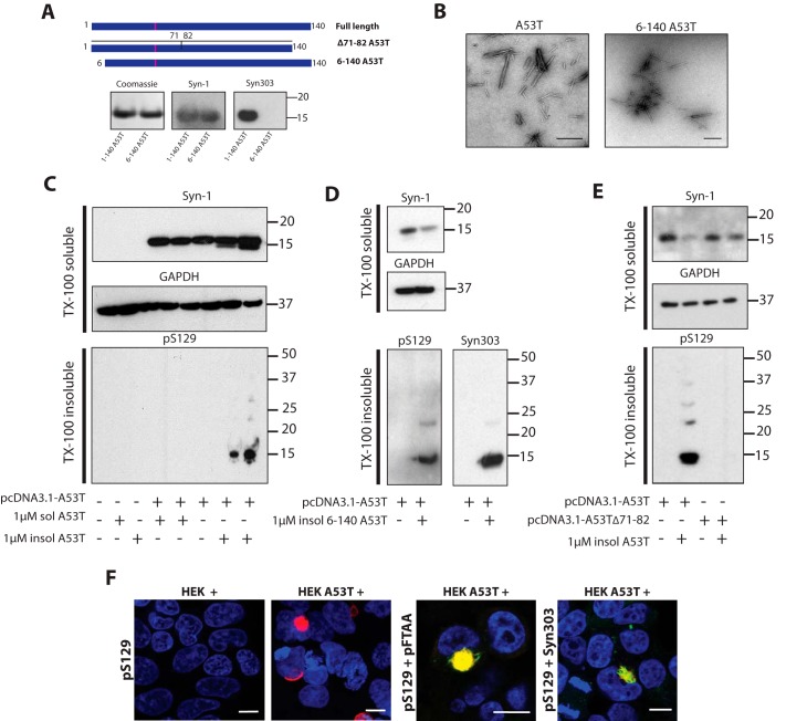
Figure 1.
Seeding assay for A53T α-synuclein in HEK 293T cells. A, full-length 1–140 and N terminally truncated 6–140 A53T α-synuclein in pRK172 were expressed in E. coli BL21 (DE3) cells. Following purification (Coomassie), immunoblotting showed that Syn-1 labeled both full-length and truncated α-synuclein, whereas Syn303 labeled full-length, but not truncated, α-synuclein. After transient transfection, full-length and Δ71–82 A53T α-synuclein were expressed in HEK 293T cells. B, electron micrographs of assembled full-length and truncated A53T α-synuclein. Scale bar, 500 nm. C, Western blot analysis of untransfected and A53T α-synuclein–transfected HEK 293T cells seeded with unassembled and assembled full-length A53T α-synuclein. Cell lysates were separated into Triton X-100–soluble and -insoluble fractions by ultracentrifugation at 100,000 × g for 1 h at 4 °C. Blots of anti-α-synuclein antibody Syn-1 and anti-GAPDH were processed for the Triton X-100–soluble fractions and anti-α-synuclein antibody pS129 was used for Triton X-100–insoluble fractions. Twenty μg of Triton X-100–soluble and 50 μg of Triton X-100–insoluble proteins were run on 4–12% BisTris SDS-PAGE. D, Western blot analysis of full-length A53T α-synuclein expressing HEK 293T cells seeded with N terminally truncated A53T α-synuclein aggregates. Triton X-100–soluble fractions were blotted with Syn-1 and anti-GAPDH. Triton X100–insoluble fractions were blotted with pS129 and Syn303. E, Western blot analysis of full-length and Δ71–82 A53T α-synuclein expressing HEK 293T cells seeded with 1 μm full-length α-synuclein aggregates. Triton X-100–soluble fractions were blotted with Syn-1 and anti-GAPDH antibodies. Triton X-100–insoluble fractions were blotted with anti-pS129 antibody. F, staining of untransfected (HEK+) and full-length A53T α-synuclein–transfected (HEK A53T+) cells seeded with 1 μm aggregated N terminally truncated 6–140 A53T α-synuclein with pS129 (red), Syn303 (green), and pFTAA (green). Nuclei were visualized with DAPI (blue). Scale bar, 10 μm.

