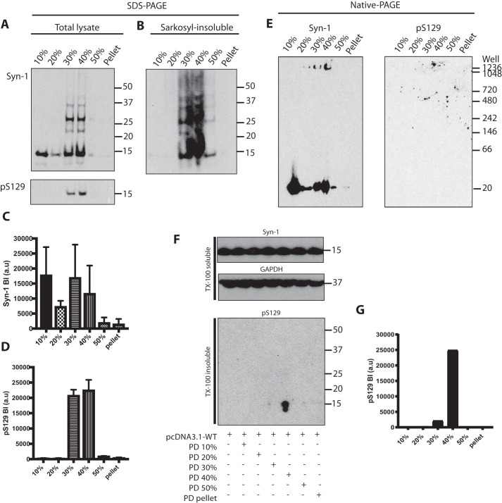
Figure 6.
Characterization of α-synuclein from Parkinson's disease brains following sucrose gradient centrifugation and identification of seed-competent species. A, Western blot analysis (anti–α-synuclein antibodies Syn-1 and pS129) following SDS-PAGE of brain lysates fractionated by sucrose gradient centrifugation. A representative blot using PD2 is shown. B, Western blot analysis (antibody Syn-1) of Sarkosyl-insoluble brain lysates. C and D, densitometric analysis of fractionated brain lysates. The results are expressed as the mean ± S.D. (n = 3). E, Western blot analysis (anti-α-synuclein antibodies Syn-1 and pS129) following native-PAGE of brain lysates fractionated by sucrose gradient centrifugation. F, Western blot analysis following SDS-PAGE of HEK 293T cells expressing human WT α-synuclein seeded with fractionated lysates of the superior temporal gyrus from an SNCA duplication case. Triton X-100–soluble cell lysates were blotted with Syn-1 and anti-GAPDH antibodies. Triton X-100 insoluble fractions were blotted with pS129. G, densitometric analysis of Triton X-100–insoluble pS129 bands of seeded cells.

