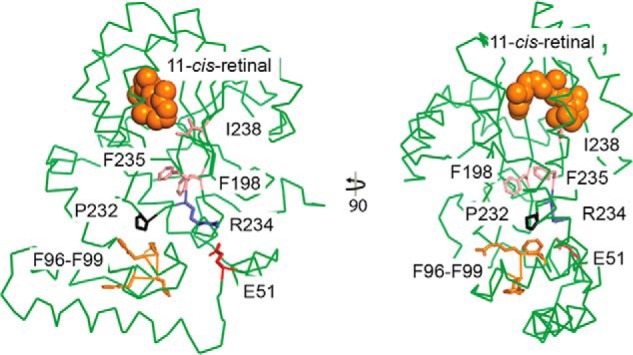
Figure 10.

Crystal structure of CRALBP. Two views of the CRALBP crystal structure (PDB 3HY5) with key residues colored as indicated. Residues in light pink (I198, F235, and I238) underwent conformational change in the R234W mutant crystal structure (PDB 3HX3, not shown) to alter 11-cis-retinal binding affinity and decreased enzymatic activity. Residues lost in the Phe96–99del mutant are shown in orange (F96, L97, R98, and F99) and the interacting Pro-232 is shown in black. 11-cis-retinal is depicted by orange spheres.
