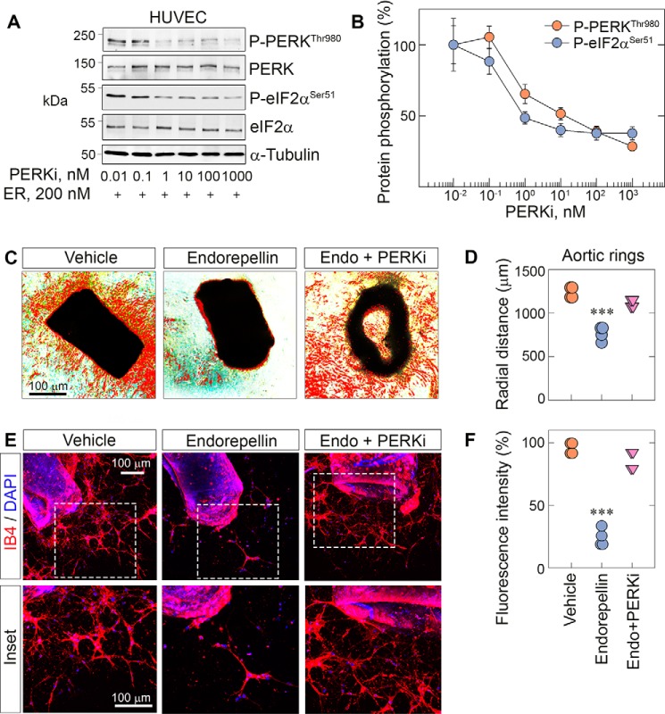
Figure 5.
PERK inhibition blocks endorepellin-dependent stress signaling and angiostasis in aortic rings. A, representative immunoblots of dose-dependent experiments of HUVECs pretreated with the PERKi for 30 min, followed by endorepellin (200 nm) exposure for 1 h and 30 min. Blots were probed for p-PERK and p-eIF2α. α-Tubulin served as the loading control. B, quantification of p-PERK and p-eIF2α from A, with normalization to their respective total protein. Data are from three independent biological experiments (n = 3), plotted as mean ± S.E. One-way ANOVA was performed on the data. C, representative phase-contrast images of aortic rings treated with endorepellin or endorepellin and the PERKi for 7 days in total after sprouting. D, quantification of the radial distance of endorepellin- or endorepellin and PERKi–treated sprouts from C. Phase-contrast data are from 12 vehicle-, 10 endorepellin-, and 10 endorepellin and PERKi–treated rings from four independent experiments (n = 4). E, representative confocal images of aortic rings treated with endorepellin or endorepellin and PERKi–treated rings. Rings were labeled with IB4 (red) and DAPI (blue). F, quantification of fluorescence intensity of the sprouted area from E. Confocal data are from eight vehicle-, eight endorepellin-, and nine endorepellin and PERKi–treated rings from four independent biological experiments (n = 4). All statistical analyses were calculated via one-way ANOVA (***, p < 0.001).

