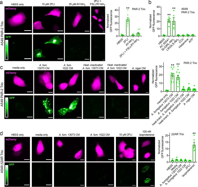
Figure 2.
Activation of PAR-2 by peptide PAR-2 agonist and A. fumigatus CM in A549 cells. Cells were transfected with the PAR-2 Trio system (26) as described in the text to detect receptor–β-arrestin interaction upon PAR-2 activation. Many other studies have already demonstrated that β-arrestin is recruited to PAR-2 upon activation (26, 29, 30, 74–78). Using this assay allows us to directly look at PAR-2 activation in the absence of any other signals downstream of other targets of proteases and/or CM that could impinge on the PAR-2 signaling pathway. a, after 90 min stimulation with PAR-2 agonist 2FLI, GFP fluorescence was increased ∼25-fold compared with buffer (HBSS) only or PAR-4–activating peptide AY-NH2. The GFP fluorescence increase, signaling PAR-2 activation, was lost when cells were stimulated with 2FLI in the presence of 10 μm PAR-2 antagonist FSLLRY-NH2. b, bar graph showing data from experiments as in a with alternative PAR-2 agonists AC 55541 and SLIGRL-NH2 plus scrambled LRGILS-NH2 and cholinergic agonist CCh, histamine, and purinergic agonist ATP (100 μm each). c, representative images of PAR-2 activation with A. fumigatus (A. fum.) CM. Cells were exposed to conditioned media for 10 min followed by washing and resuspension in HBSS for 90 min. Bar graph on right shows normalized GFP fluorescence. Significance was determined by one-way ANOVA with Dunnett's post-test comparing values to media only control; **, p < 0.01. d, A549 cells were transfected with β2AR Trio. A. fumigatus CM did not activate β2AR but 100 nm isoproterenol did. Bar graph shows quantification of results. All bars show data points from ≥7 independent experiments imaging fields from independent transfected wells on separate days. Significance was determined by one-way ANOVA with Dunnett's post-test comparing all values to HBSS alone; **, p < 0.01. Scale bars are 15 μm.

