Figure 5.
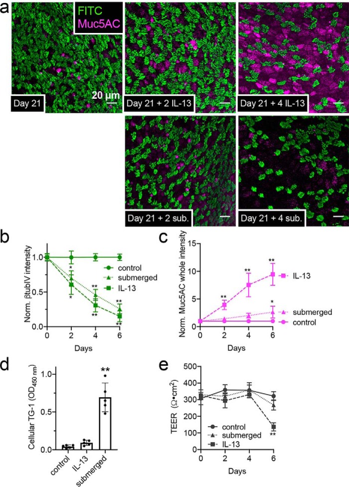
Loss of cilia with IL-13 treatment or submersion of ALI cultures of primary human nasal cells. a, representative immunofluorescence (IF) of cilia (β-tubulin IV; green) and Muc5AC (magenta) in ALIs 21 days after air (top left). Top, middle, and right show cilia loss after further 2–4 days basolateral IL-13. Bottom, middle, and right show loss of cilia with less Muc5AC with apical submersion. b and c, cilia loss (b; normalized β-tubulin IV IF) and Muc5AC increase (c) in cultures (starting at day 21) exposed to subsequent IL-13, apical submersion, or no treatment (control). Ten fields from one ALI from one patient were imaged and averaged for an independent experiment; results shown are mean ± S.E. of 4–6 independent experiments (4–6 patients). d, squamous marker TG-1 quantified by ELISA at day 25 (21 days at air and then 4 subsequent days of IL-13, submersion, or no treatment). Each data point is an independent ALI from a different patient (n = 5 total ALIs). Significance was determined by one-way ANOVA with Dunnett's post-test; **, p < 0.01. e, TEER at time points as in b and c. Significance was determined by one-way ANOVA with Bonferroni post-test; *, p < 0.05, and **, p < 0.01.

