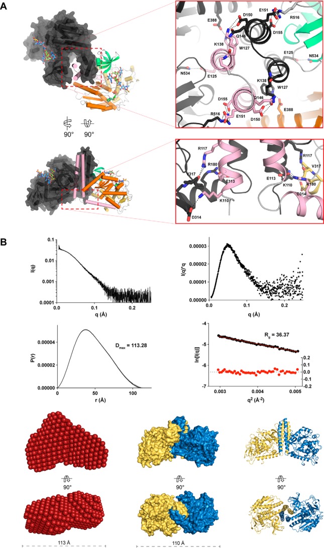
Figure 4.
Interactions between the N-terminal coiled-coil domains drive FUT8 dimerization in solution. A, self-association of the N-terminal helices of FUT8 creates a four helix bundle that buries hydrophobic residues and creates multiple interchain salt bridges. B, intensity plot of FUT8 SAXS scattering (top left), a Kratky plot derived from FUT8 scattering showing that it forms a compact particle in solution (top right), and P(r) and Guinier plots indicating that FUT8 has a maximum dimension of 113.28 Å in solution (bottom left) and a radius of gyration of 36.37 Å (bottom right). C, a bead model of FUT8 in solution generated from solution scattering data (left) corresponds well with the dimer observed in the FUT8 crystal structure, shown as a surface (center) and cartoon (right) view.

