
Figure 6.
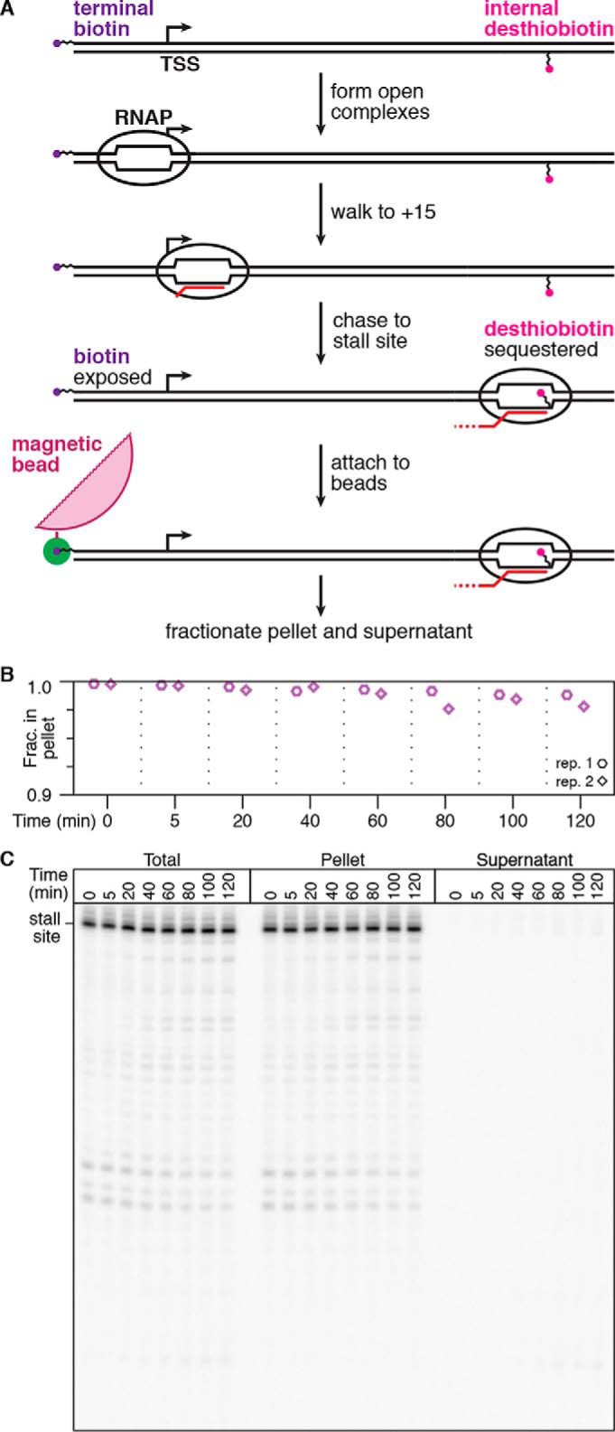
Quantification of elongation complex stability at a desthiobiotin–TEG stall site. A, overview of the experiment shown in B and C. Using a DNA template with an internal desthiobiotin–TEG stall site and a 5′-terminal biotin, RNAP is positioned at the stall site to sequester desthiobiotin, but the 5′-biotin remains exposed. These complexes are immobilized on streptavidin-coated magnetic beads, washed to remove free NTPs, and incubated at room temperature before separation into bead pellet and supernatant fractions. B, quantification of the fraction of desthiobiotin–TEG–stalled TECs retained in the bead pellet over time. The y axis range is 0.9–1.0 to facilitate clear data visualization. C, gel showing replicate 1 from B. Pellet and supernatant data points in B and C are from two independent replicates; the total reaction control was performed once.