Figure 5.
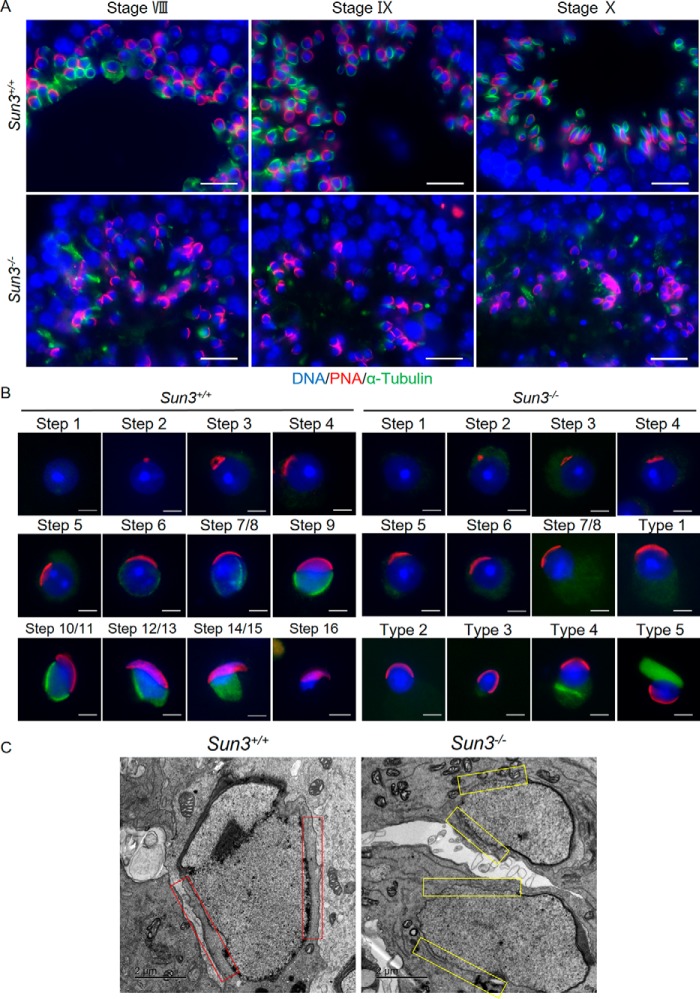
Manchette formation is disrupted in Sun3−/− mice. A, representative images of testis sections from 8-week-old Sun3+/+ and Sun3−/− mice, stained by anti-PNA (red) and anti-α-tubulin (green) antibodies. DNA was counterstained with Hoechst. Scale bars = 20 μm. B, representative images of spermatids from 8-week-old Sun3+/+ and Sun3−/− mice, stained by anti-PNA (red) and anti-α-tubulin (green) antibodies. DNA was counterstained with Hoechst. Types 1–5 show various abnormal spermatids in Sun3−/− mice. Scale bars = 2 μm. C, manchette microtubule bundles were not observed in Sun3−/− mice by transmission EM. Red rectangles show the presence of manchette microtubule bundles in Sun3+/+ mice, whereas yellow rectangles indicate the absence of such microtubule bundles in Sun3−/− mice. Scale bars = 2 μm.

