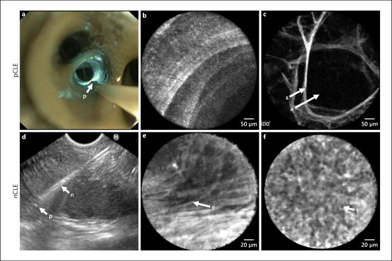
Fig. 4.
pCLE imaging procedures in vivo with corresponding images of the normal alveolar and airway wall compartment (a–c); nCLE imaging in vivo with corresponding images of reactive lymph node capsula and cortex (d–f). a pCLE with the probe (p) positioned in the central airways of the right lower lobe on its way to be advanced to the alveolar compartment. 488 nm laser light reflecting at the airway wall (blue). b pCLE image of the distal airway wall showing a helical ring-like pattern of the terminal bronchiole. c pCLE image of the alveolar compartment showing air-filled alveoli (a) and alveolar septae (s). d nCLE during an EUS procedure for staging of lung cancer, with the probe (p) extending 2 mm distal to the tip of the needle (n). e nCLE image showing elastin fibres (e) of the capsula of a lymph node. f nCLE image showing lymphocytes (l) in a reactive lymph node Wijmans et al. [85]. pCLE, probe based confocal laser endomicroscopy; S, septae; A, alveolar space; nCLE, needle based confocal laser endomicroscopy.

