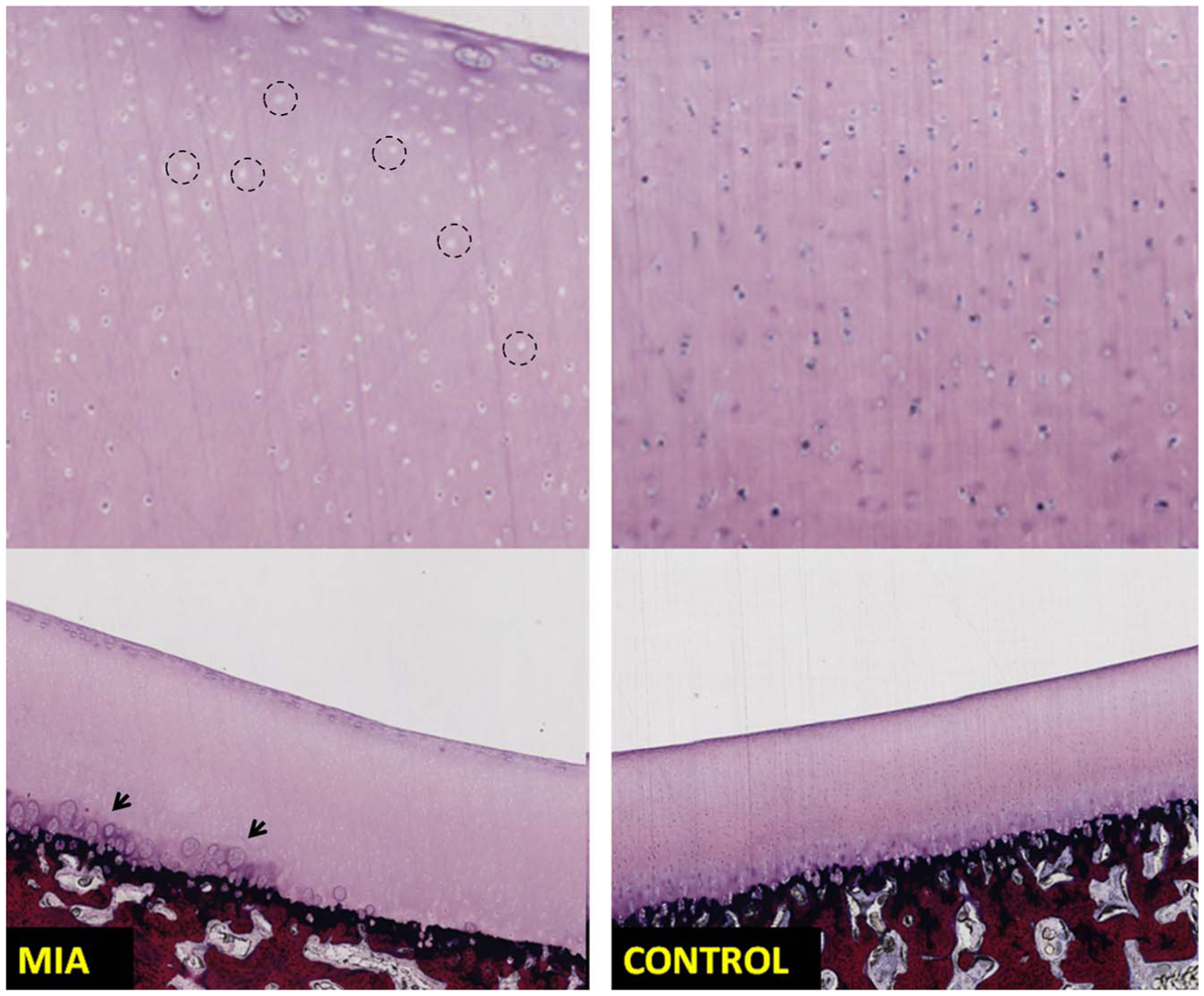
FIGURE 5.

Sections of patella stained using methylene blue/basic fuchsin show chondrocytes loss (left top image, example regions are marked using circles) in the MIA injected knee (left column) compared with the control knee (right column) exhibiting normal chondrocyte appearance. Chondrocyte clustering and degeneration (marked using arrows) was noticed in the MIA knee. The MIA knee from the same swine was marked as grade 1 (cartilage heterogeneity) on the PCD-CT images.
