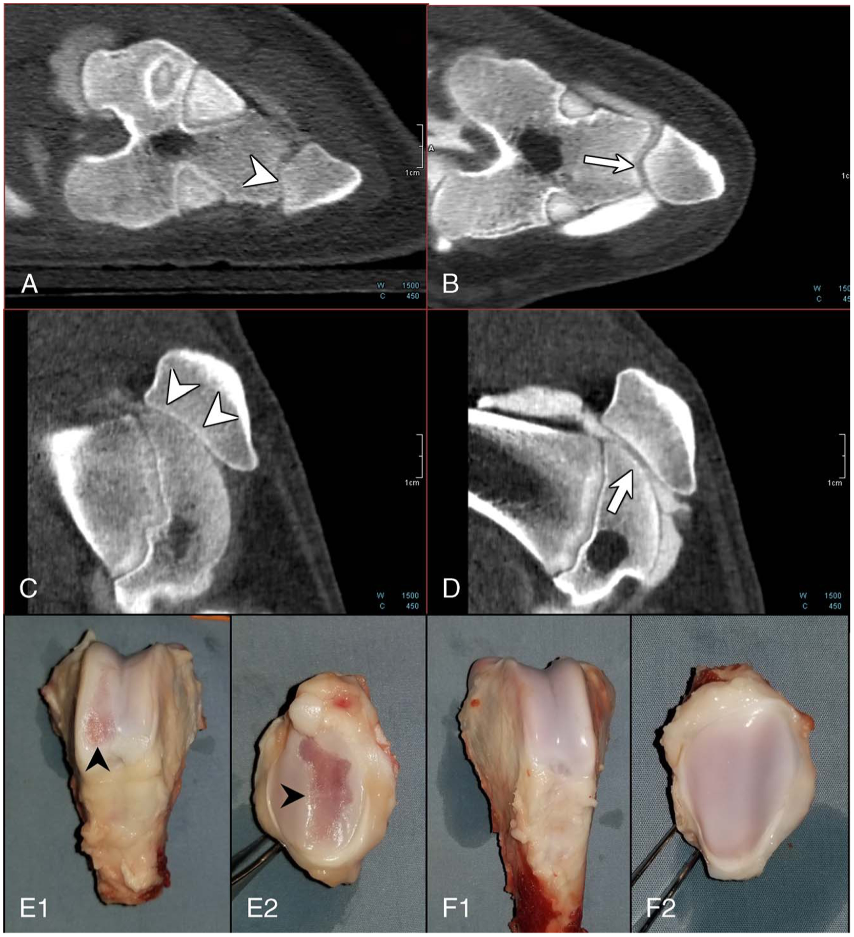
FIGURE 6.

Photon-counting detector computed tomography (PCD-CT) images and photographs of harvested knees showing substantial cartilage loss from the articular surface. PCD-CT images from the MIA knee (A and C) show bone-on-bone articulation (arrowheads in A, C) due to complete cartilage loss (grade 3), whereas the control knee showed normal cartilage thickness and appearance (grade 0). Excised femur (E1, F1) and patella (E2, F2) specimens from the same pig showed substantial cartilage damage (black arrowheads) in the MIA knee (E1, E2) compared with the control knee, which exhibited normal cartilage appearance (F1, F2).
