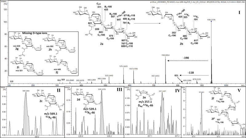
Fig 7: MS2 fragmentation of succinylated Man1Ara5.
(m/z 939 (M-H)1−; 2a) identifying dominant linear arrangement of Ara5 termini. I) MS2 profile of 2a and representative fragmentations. II, III & IV) Fragment ions confirming the location of the succinyl residue at the 3-position of 2-glycosylated Araf. V) The ion at m/z 557 corresponding to structures 2f and 2f’ represents the possibility of Ara5-skeleton being linear or branched. Inset) Missing ions (2g-j) would have confirmed a branched structure.

