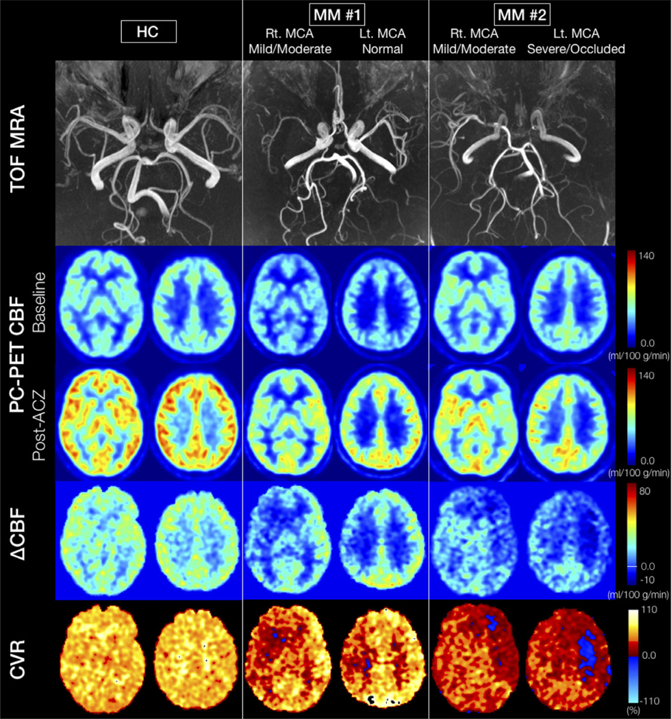
FIGURE 5:

Representative cases: (From the top row) TOF MR angiogram, two slices of PC-PET CBF at baseline and after ACZ administration, change in CBF (ΔCBF), and cerebrovascular reactivity (CVR, %) maps. The left column shows an HC case. A significant CBF increase is visible in all territories after ACZ. The ΔCBF in GM is larger than deep WM, while CVR is essentially constant in all regions for the HC. The middle and right columns show the patients with Moyamoya disease. Both cases present asymmetrical stenosis grades for each MCA with normal grades for the bilateral PCAs. Bilateral ACAs are graded as normal in Patient #1 and as severe in Patient #2. The post-ACZ CBF is obviously asymmetrical, with less CBF increase on the hemisphere with more severe artery stenosis in both MM cases. In particular, in the left hemisphere of Patient #2, there are regions where the CVR is negative (blue area). HC: healthy control, TOF: time of flight, ACZ: acetazolamide, PC: phase-contrast, PET: positron emission tomography, CBF: cerebral blood flow, CVR: cerebrovascular reactivity, MCA: middle cerebral artery, PCA: posterior cerebral artery, ACA: anterior cerebral artery.
