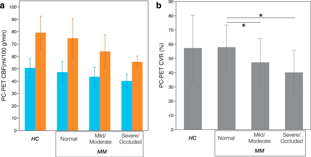
FIGURE 6:
PC-PET CBF (a) and CVR (b) was compared by MR angiogram stenosis grades (for MM patients) as well as for HC for ASPECTS cortical regions. No differences in baseline CBF were identified between grades of stenosis. However, regions in Moyamoya patients corresponding to mild/moderately stenosis and severe/occluded arteries showed lower CBF than in normal regions. Blue and orange bars in (a) show the CBF of baseline and post-ACZ scan, respectively. The error bars show standard deviation. Asterisks indicate significant difference at the P < 0.05 level. HC: healthy control, MM: patients with Moyamoya disease, PC: phase-contrast, PET: positron emission tomography, CBF: cerebral blood flow, CVR: cerebrovascular reactivity.

