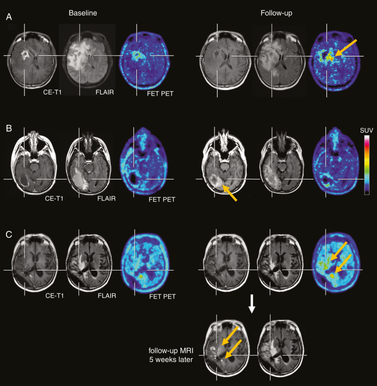
Figure 1.
(A, top row) The contrast-enhanced MRI, the FLAIR-weighted MR image and FET PET at baseline (left three images) and after two cycles of regorafenib (right three images) of a 35-year-old glioblastoma patient (IDH-wildtype). Following regorafenib, the MRI showed a “Partial Response” according to RANO criteria, whereas the FET PET depicted increased metabolic activity spatially located medial to the area with the necrotic core on MRI (arrow), indicating pseudoresponse on MRI. The patient deceased 3 months later. (B, second row) Discrepant MRI and FET PET findings in a 39-year-old glioblastoma patient (IDH-wildtype), which indicate pseudoprogression on MRI. Due to a distant tumor recurrence (right parietal, images not shown), the patient was treated with regorafenib. After two cycles of regorafenib, the MRI showed an increase of both the FLAIR signal alteration and contrast enhancement at the medial edge of the initial resection cavity (arrow, second row), whereas the FET PET revealed no pathologically increased tracer uptake. Despite a clinical stable course for the next three months, the patient died unexpectedly most probably due to a progression of the distant tumor. (C) Pathological increase of FET uptake after two cycles of regorafenib (arrows, third row), whereas the corresponding MRI is consistent with “Stable Disease” according to RANO criteria. Five weeks later, the 43-year-old patient deteriorated clinically, and the MRI showed a progression of the contrast-enhancing lesions and of the FLAIR signal (arrows, bottom row).

