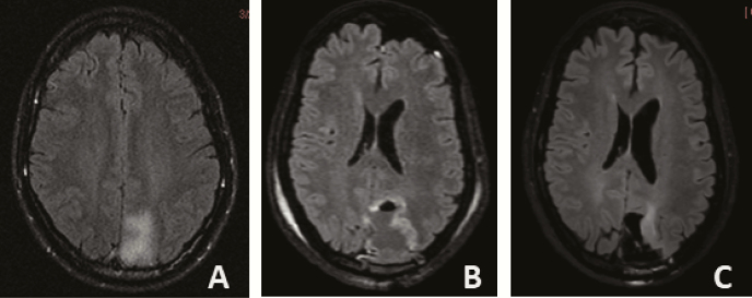
Figure 1.
Axial T2/FLAIR MRI images of patient with left occipitoparietal oligodendroglioma WHO grade 2, IDH1 mutated, 1p19q codeleted with indolent course. (A) Incidental finding at age 39, followed with serial imaging without intervention. (B) Near total resection at age 41 after slight increase in T2/FLAIR hyperintensity and development of headaches, then followed without additional treatment. (C) Slight increase in T2/FLAIR hyperintensity adjacent to the surgical cavity at age 52 (13 years from initial imaging diagnosis; no prior radiation or chemotherapy).

