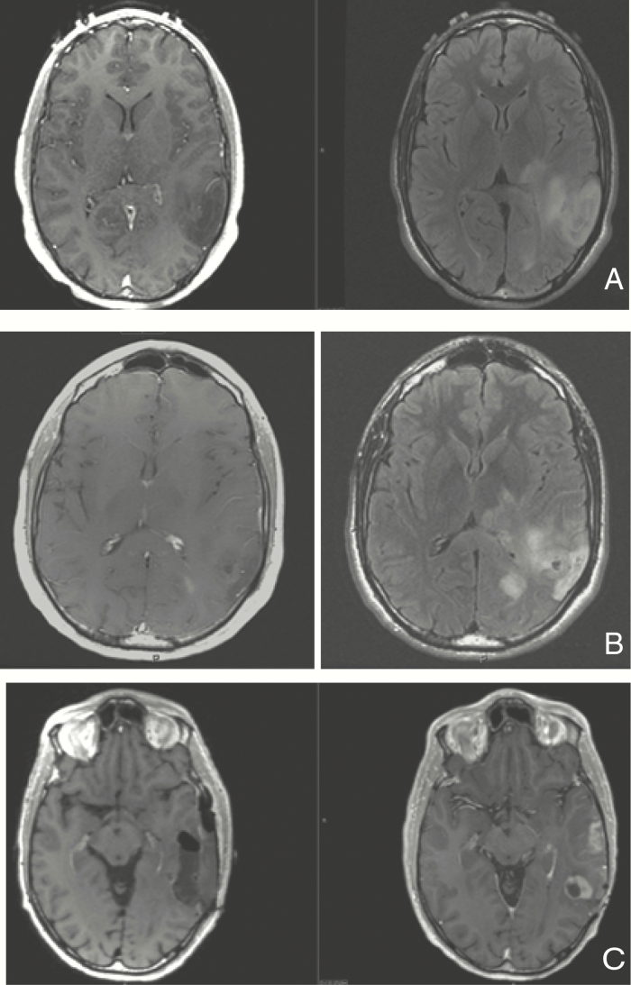
Figure 2.
Axial T1 with contrast and T2/FLAIR MRI images of patient with left posterior temporal oligodendroglioma WHO grade 2, IDH1 mutated, 1p19q codeleted with aggressive course. (A) Imaging at age 28 after presenting with seizures; underwent biopsy without further treatment. (B) Imaging at age 31 after biopsy demonstrating transformation to anaplastic oligodendroglioma. Patient had received 2 years of therapy with temozolomide due to imaging progression detected 6 months after initial diagnosis. (C) Imaging preresection (right) and postresection (left) at the time of 4th progression (age 37), after having received radiation, procarbazine and lomustine; received subsequent treatment with re-irradiation and with a PD-1 inhibitor 3 years later for new recurrence.

