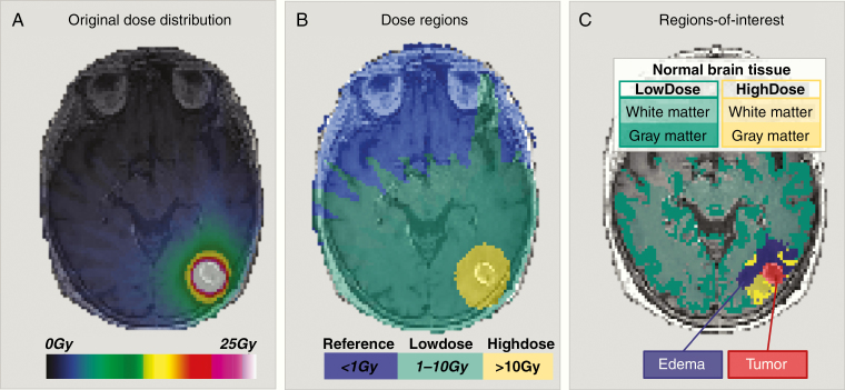
Figure 1.
Regions-of-interests used for assessment of normal-appearing brain tissue responses to SRS. (A) A representative co-registered stereotactic dose distribution for a patient with brain metastasis from non-small cell lung cancer as an overlay of a T1-weighted post-contrast image acquired pre-SRS. The prescribed SRS dose was 25 Gy. (B) The dose distribution was divided into 3 dose regions: Reference: <1 Gy (blue overlay), LowDose: 1–10 Gy (green overlay), and HighDose: >10 Gy (yellow overlay). (C) The final regions-of-interest used for longitudinal response assessments included normal-appearing brain tissue, ie, white and gray matter in LowDose (white matter: light green overlay, gray matter: dark green overlay) and HighDose (white matter: light yellow overlay, gray matter: dark yellow overlay), excluding edema (purple overlay) and tumor (red overlay).

