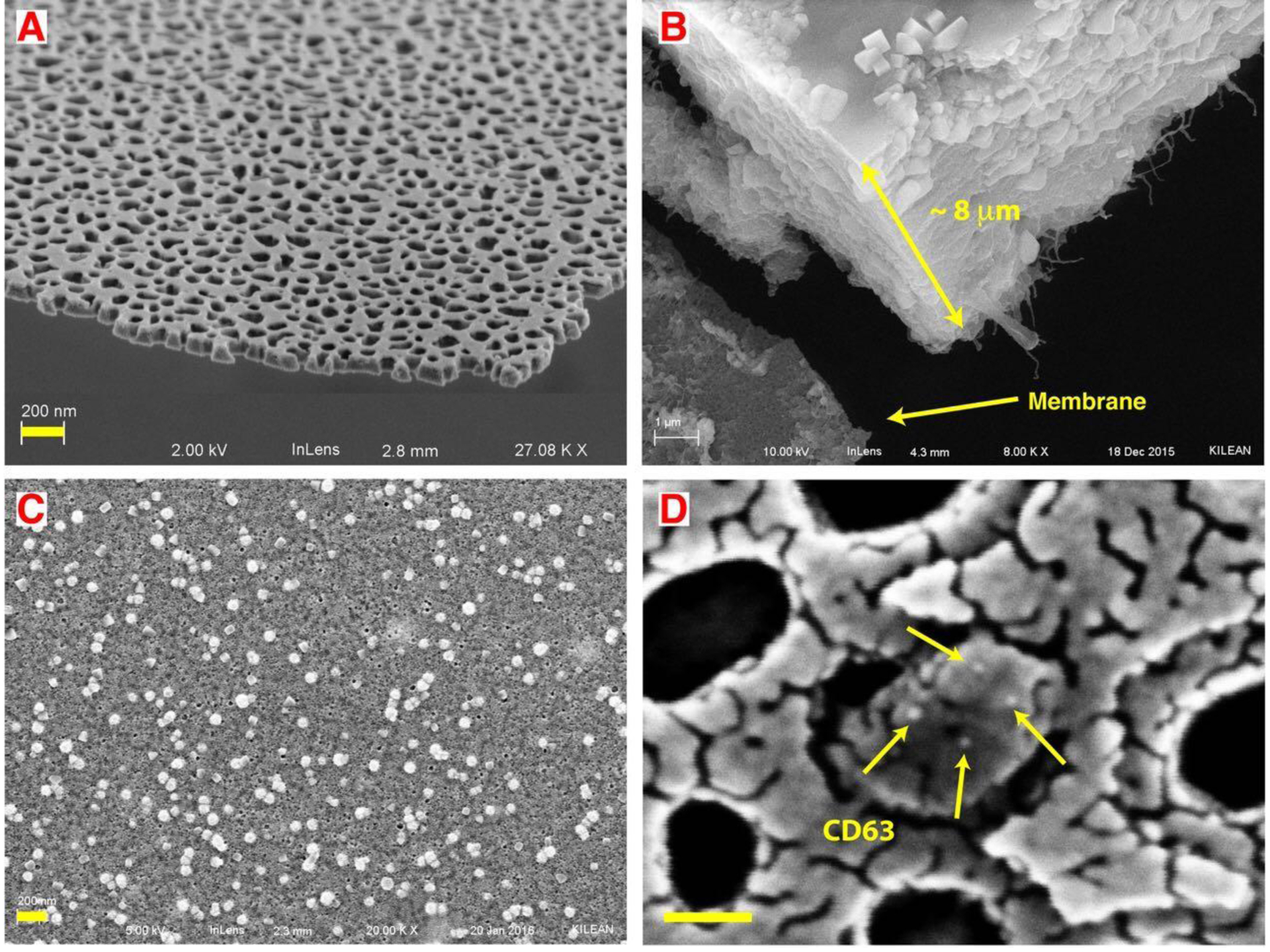
Figure 2: Small extracellular vesicles (sEV) captured from undiluted blood plasma.

A) SEM images showing the thinness and high porosity of nanoporous silicon nitride (NPN). B) In normal flow filtration (NFF) a protein cake of ~8 μm cake rapidly builds up on the membrane surface. C) After capturing and cleaning steps of TFAC, small vesicles are captured on the membrane surface with minimum fouling. D) Nanogold conjugated anti-CD63 antibody labels an EV captured in a pore multiple times, indicating it is likely a CD63 positive sEV. Note: the fragmented appearance of the surface results from the use of a limited amount of gold (3 nm) to avoid obscuring the gold label on the antibody (18 nm). By contrast 10 nm of gold was sputtered on the samples to avoid charging effects in SEM in both B and C. Scale bar = 200 nm for A, B and C. Scale bar = 50 nm for D.
