
Fig. 1. MMP9 is locally activated in epithelial cells as a result of direct contact with fibroblasts.
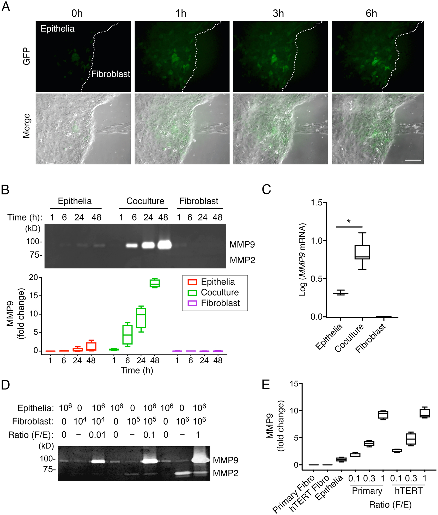
(A) Time-lapse phase-contrast and fluorescence images of immortalized human corneal epithelial cells carrying the MMP9-eGFP reporter construct and primary human corneal fibroblasts. (B) Gelatin zymography of culture media from epithelial cells and fibroblasts grown alone or in a mixed coculture system, in time course experiments. n = 4 independent experiments. (C) qPCR analysis of MMP9 expression in cells grown alone or in coculture for 24 h. n = 8 independent experiments. (D) Gelatin zymography showing MMP activity with increasing numbers of fibroblasts relative to epithelial cells in coculture for 24 h. n = 3 independent experiments. (E) Gelatin zymography of culture media from cells grown alone or in coculture using immortalized human corneal epithelial cells and either primary human corneal fibroblasts or telomerase-immortalized human corneal fibroblasts (hTERT). n = 4 independent experiments. The box and whisker plots show the 25 and 75 percentiles (box), the median (horizontal line in box), and the minimum and maximum data values (whiskers). Significance was determined using the Wilcoxon test. *, p<0.05. Scale bar, 250 μm.