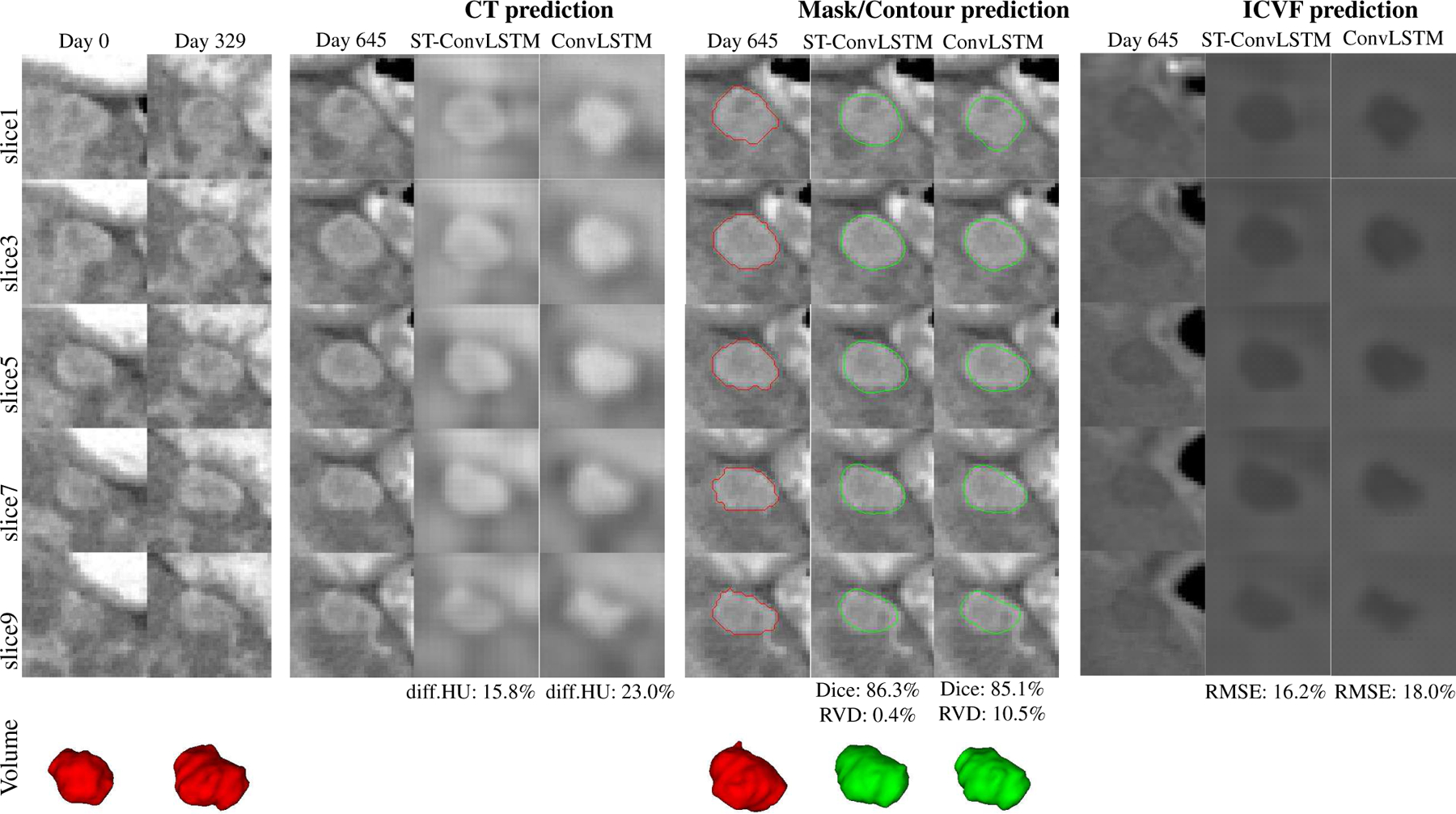
Fig. 5.

An illustrated example shows the prediction results of CT, mask/volume, and ICVF of a tumor by ST-ConvLSTM and ConvLSTM. Note that the tumor contours are superimposed on the ground truth CT images at time 3. Red: ground truth boundaries; Green: predicted tumor boundaries. In this example, consecutive image slices with the spatial interval of two slices are shown for better visualization of the spatial changes/differences.
