
Figure 1.
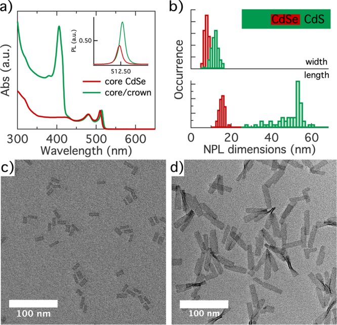
(a) Absorption spectrum of the initial CdSe core and final CdSe/CdS core/crown nanoplatelets (NPLs), normalized at the maximum of the heavy-hole absorption line. The inset shows the photoluminescence (PL) spectra of core and core/crown NPLs, with the area normalized to the respective PLQY. (b) Histograms of (top) the width and (bottom) the length of (red) the initial core and (green) the final core/crown NPLs. The inset shows a scheme representing the average heterostructure to scale. (c) Overview bright field TEM image of initial CdSe crown NPLs. (d) Same as panel c for the final CdSe/CdS core/crown NPLs.