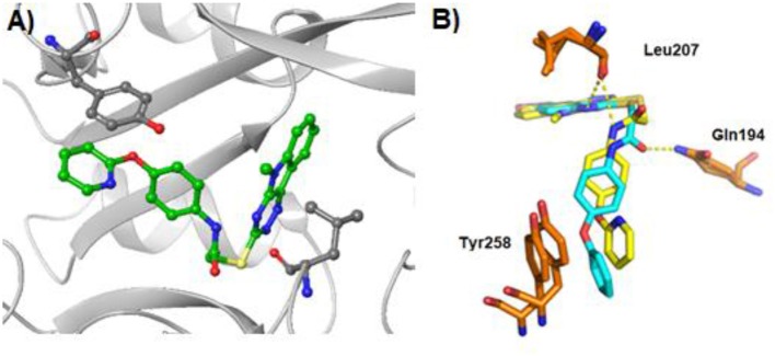
Figure 3.
X-ray crystal structure of PqsR in complex with compound 40 and M64 (PDB:6B8A) (Kitao et al., 2018) (A) X-ray co-crystal structure of 40 bound to PqsR LBD. The protein structure is presented in gray and residues Tyr258 and Leu207 are labeled in black. (B) Overlapping crystal structures of 40 and M64 in complex with PqsR ligand binding domain. Compound 40 is represented in yellow and M64 in light blue.

