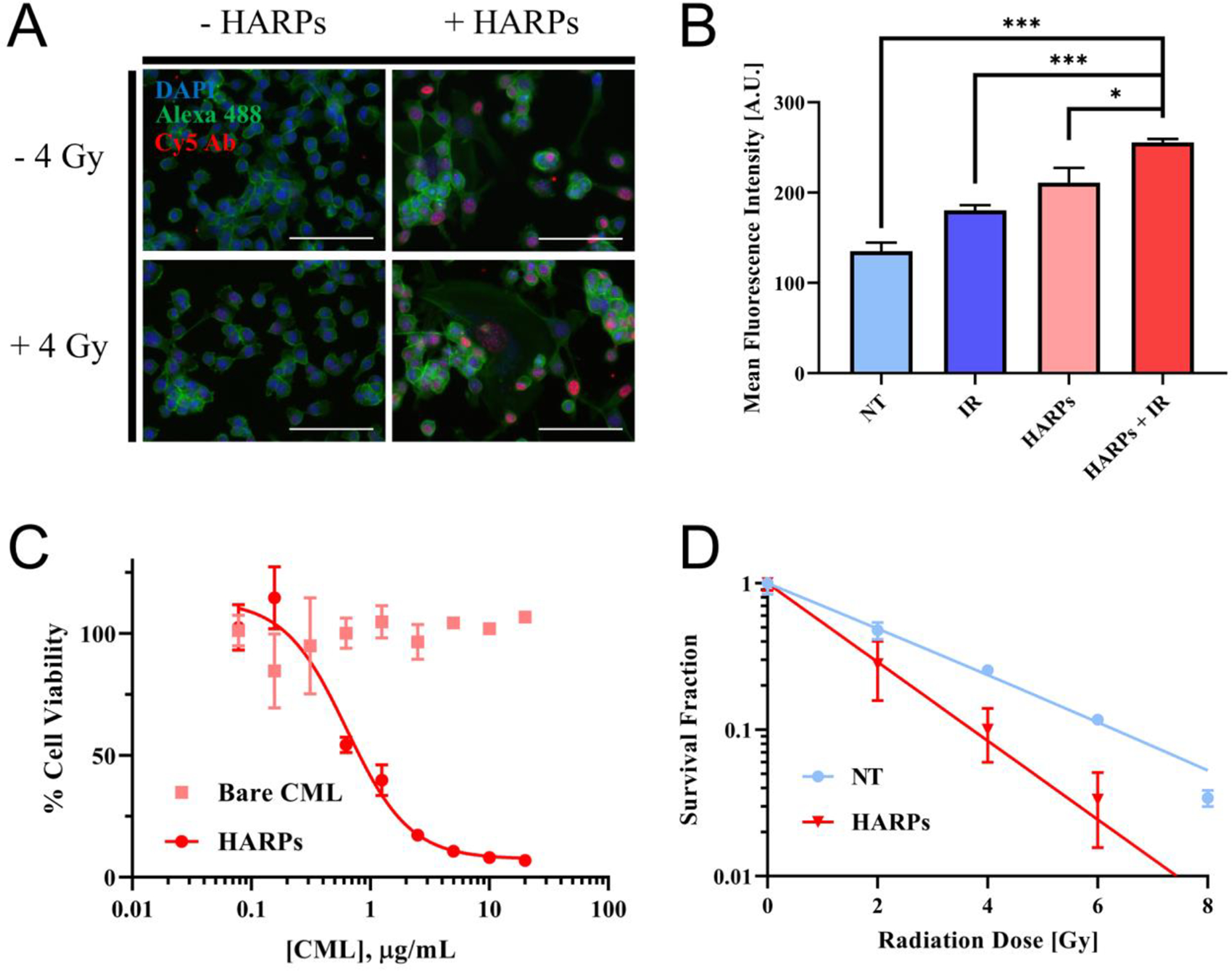
Figure 4.

In vitro efficacy of HARP and combination therapy. (A) Fluorescence microscopy of γH2AX assay. IR (4 Gy), HARPs (24 hour incubation, 0.55 μg/mL RTX), and a combination shows brighter fluorescence, indicating DNA damage at these “foci”. Scale bar = 100 μm. (B) Mean fluorescence intensity measured by flow cytometry of 105 cells (in triplicate for each group, mean ± standard deviation shown) shows an increase in DNA damage when either IR or HARPs are used, with further damage from the combination of the two treatments. Data significance evaluated with unpaired student t-test, *, P < 0.05, **, P < 0.01, ***, P < 0.001. (C) Cell viability at 72 hours shows little to no toxicity for bare nanoparticles, with a large increase in toxicity as HARPs approaches 1 μg/mL. (D) Survival fractions show reduced clonogenicity of cells treated with 0.55 μg/mL HARPs for 24 hours before exposure to IR (compared to those exposed to IR alone).
