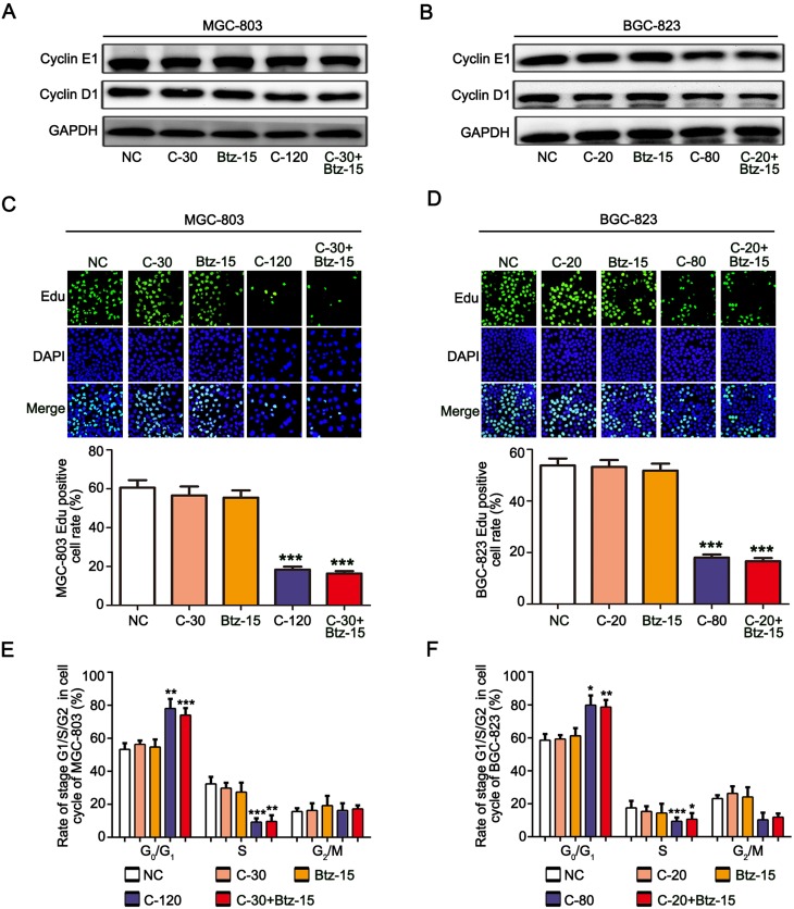
Figure 2.
Chidamide combined with bortezomib inhibited the proliferation of the MGC-803 and BGC-823 cell lines. The MGC-803 and BGC-823 cell lines were treated with chidamide (30 µM for MGC-803 or 20 µM for BGC-823), bortezomib (15 nM), chidamide (120 µM for MGC-803 or 80 µM for BGC-823), or chidamide (30 µM for MGC-803 or 20 µM for BGC-823) in combination with bortezomib (15 nM) for 48 hours. (A and B) Representative images of cyclin D1 and cyclin E1 expression. (C and D) EdU staining, showing cells in a proliferating state after the different treatments, and their statistical results. (E and F) Cell-cycle distribution of MGC-803 and EC-109 cells were confirmed after the different treatments by propidium iodide staining and FACS analysis. (One-way ANOVA with Bonferroni’s post hoc test was applied to compare the indicated groups. *P<0.05, **P<0.01, and ***P<0.001, compared with the negative control, chidamide-, or bortezomib-alone groups). The experiment was repeated at least three times. Negative control: DMSO.

