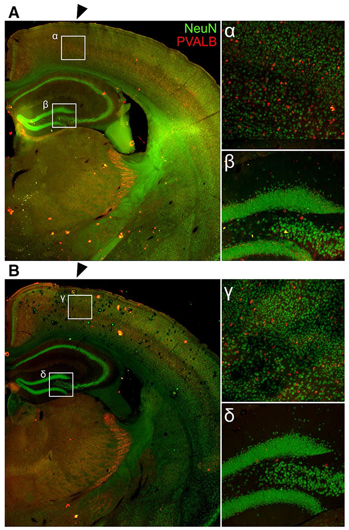
Fig. 2.

Healing at the cortical impact site. There were no differences after 28 d of healing at the cortical impact site (black arrows) in number of NeuN + neurons, PVALB + neurons, or PNNs in TBI + MM Control (a) or TBI + 181a inhibitor (b). Magnified views of the cortex just below the impact site (α,γ) and the dentate gyrus/CA3c (β,δ)
