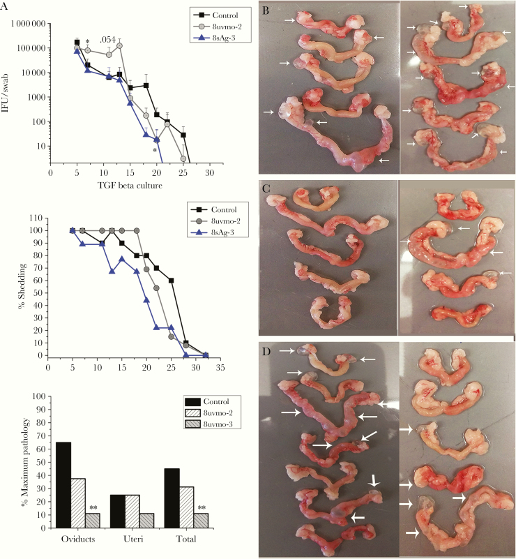
Figure 3.
Adoptive transfer of T-cell clones maintained in transforming growth factor (TGF)β cultures. Medroxyprogesterone-treated C57BL/6 mice received phosphate-buffered saline (control) or 1 million CD8 clone cells, 8uvmo-2 (Tc1) or 8sAg-3 (CD8γ13), 24 hours before vaginal challenge with 5 × 10⁴ inclusion forming units (IFU) Chlamydia muridarum. (A) Shedding as IFU/swab, percentage of mice shedding over time, and immunopathology score on day 56. Genital tracts from (B) control mice, (C) 8sAg-3 mice, and (D) 8uvmo-2 mice. White arrows indicate damage to uterine horn or hydrosalpinx. Data are from 2 independent experiments; number of mice = number of genital tracts. Shedding was analyzed with Dunnett’s test, and pathology was analyzed with Fisher’s exact test; *, P < .05 and **, P < .005.

