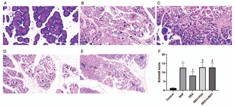
Figure 2.

Effects of dexmedetomidine on pancreatic histopathological changes. Pancreatic tissue sections were stained with hematoxylin and eosin (Original magnification ×400) at 6 h (A–E). (A) Control group; (B) SAP group; (C) DEX group; (D) DEX + VGX group; (E) DEX + α-BGT group. (F) Histopathologic severity scores of pancreatic injury. ∗P < 0.05 vs. Control group; †P < 0.05 vs. SAP group; ‡P < 0.05 vs. DEX group. The data presented are the mean ± standard deviation (n = 8). Control group: animals received a sham operation; SAP group: animals received induction of severe acute pancreatitis (SAP); DEX group: animals received a 30 μg/kg intraperitoneal injection of dexmedetomidine (DEX) 30 min before induction of the SAP model; DEX + VGX group: animals were subjected to right cervical vagotomy followed by exactly the same procedures as the DEX group; DEX + α-BGT group: animals received the selective α7nAchR inhibitor α-bungarotoxin by intraperitoneal injection (1 μg/kg) 30 min before DEX followed by exactly the same procedures as the DEX group.
