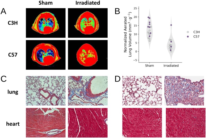
Fig 3. Radiation-induced lung injury was observed in C3H and C57 mice.
(A) CT images of the mouse thorax pseudo-colored based on radio-opacity, with the color grading from blue to red representing increased opacity. Using radio-opacity to approximate tissue density, aerated lung volume (blue) is reduced in irradiated animals compared to controls. (B) Quantification of the aerated lung volume, normalized by the animal weight, show that there was a statistically significant reduction in aerated lung volume in irradiated animals (p-value < 0.05). (C and D) Histological sections of C3H (C) and C57Bl/6 (D) heart and lung tissue stained with Masson’s trichrome.

