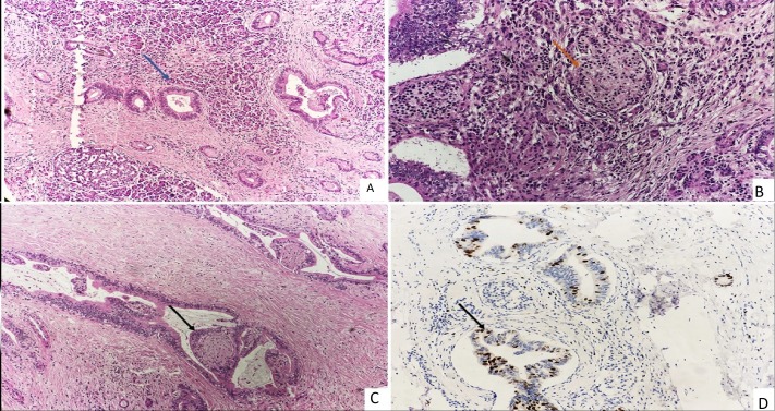
Figure 1.
(A) Unremarkable acini admixed with atypical duct-like structure (blue arrow) (H&E, 10×). (B) Clusters of neuroendocrine cells (orange arrow) along with acini (H&E, 20×). (C) Invasion of perineural space (black arrow) (H&E, 10×). (D) Ki 67 is 40% in malignant ductal structures (black arrow) and less than 2% in neuroendocrine cells (Immunohistochemistry, 20×).

