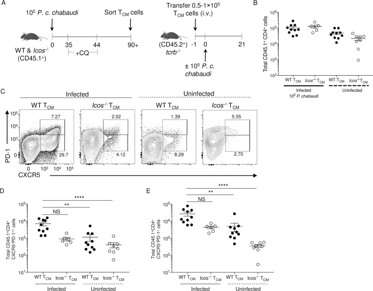
Fig 7. Icos deficiency restricts TCM cells from producing Tfh effector cells after reactivation.
(A) Experimental model. WT and Icos-/- CD45.1+ mice were infected with 105 P. c. chabaudi pRBCs and given CQ beginning at day 35 p.i. TCM cells were sorted from WT and Icos-/- CD45.1+ mice on day 90. 50–100,000 cells of each TCM cell population were transferred retro-orbitally into separate CD45.2+ tcrb-/- mice. WT CD45.2+ and tcrb-/- mice that did not receive donor cells served as controls. Twenty-four hours later, half the mice were infected with 105 P. c. chabaudi pRBCs. Mice were sacrificed at day 21 p.i. (B) Total number of live CD45.1+ CD4+ T cells recovered on day 21 p.i. (day 22 post-transfer). (C) Representative plots of CXCR5 and PD-1 expression on live activated (CD44hiCD62Llo) CD45.1+ CD4+ T cells at day 21 p.i. Upper gates indicated GC Tfh (CXCR5+PD-1++) cells, and lower gates represent Tfh-like (CXCR5+PD-1+) cells. The total number of live activated CD45.1+ CD4+ (D) GC Tfh and (E) Tfh-like cells. Data are pooled from two independent experiments with at least three mice per group (error bars, s.e.m.). Significance calculated by one-way ANOVA Kruskal-Wallis test with post hoc Dunn’s multiple comparisons test. ** p < 0.01, **** p < 0.0001, NS not significant.

