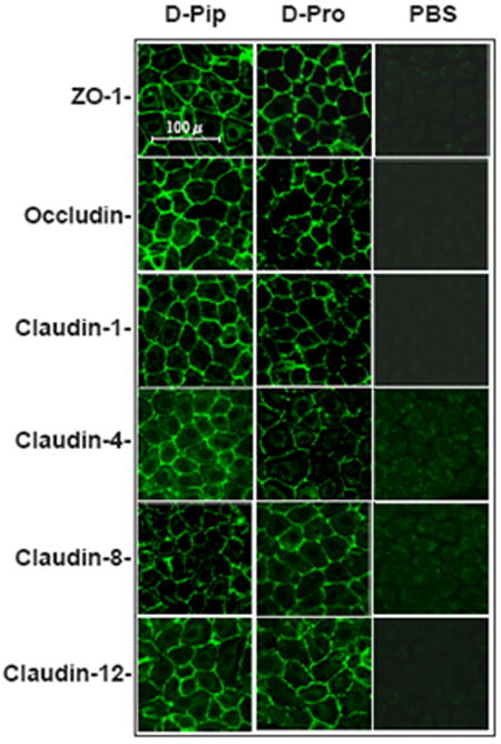
Figure 6. Immunofluorescence confocal microscopy of IC/PBS cell explants treated with d-pipecolic acid as-APF, d-proline as-APF, or vehicle (PBS) alone for 9 days.

Explanted cells from an IC/PBS donor were treated with 2.5 μM d-proline as-APF, d-pipecolic acid as-APF, or diluent (PBS) control twice weekly. On day 9, cells were fixed with acetone/ethanol and incubated with FITC-labeled anti-ZO-1 antibody, or primary antibodies against claudin or occludin proteins followed by FITC-labeled secondary antibodies. Images were obtained on a Zeiss LSM510 confocal laser scanning microscope. (Figure is representative of three separate experiments using cells from 3 IC/PBS donors)
