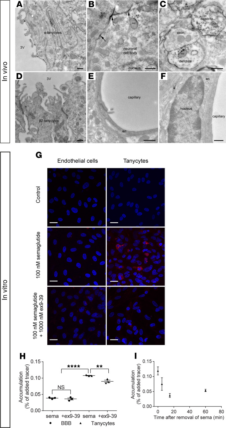
Figure 4. GLP-1R is present on tanycytes but not endothelial cells in rat ARH, and in vitro, semaglutide does not interact with BBB endothelial cells but is taken up by tanycytes.
Electron micrographs of rat tissue section from the ARH showing GLP-1R immunoreactivity (silver grains) on (A) the ventricular surface and interwoven lateral surface of α tanycytes, (B) the cell membrane of neuronal perikarya (arrows), and (C) the cytoplasm and surface of dendrites. (D) Limited, scattered GLP-1R immunoreactivity in β2 tanycytes lining the ventricular wall of the ME. (E and F) Endothelial cells lining capillaries in the ARH appear unlabeled. (G) SemaglutideCy3 (100 nM; in red) uptake in bovine brain endothelial cells (left column) in coculture with rat astrocytes and in rat tanycytes in monoculture (right column), with or without 1000 nM exendin 9-39 (ex-9-39). Nuclei Hoechst staining (blue). (H) Intracellular accumulation of 125I-semaglutide (0.7 nM) in BBB endothelial cells and tanycytes with or without 1000 nM ex-9-39. Individual values, mean, and SD shown (n = 3). Means were compared using 2-way ANOVA with Bonferroni’s correction. **P < 0.01, and ****P < 0.0001. (I) Intracellular accumulation of 125I-semaglutide in preloaded tanycytes in clean uptake buffer. Data are shown as mean and SD (n = 3). Scale bars: 500 nm (A–F), 25 μm (G). en, endothelial cell; 3V, third ventricle; mit, mitochondrion.

