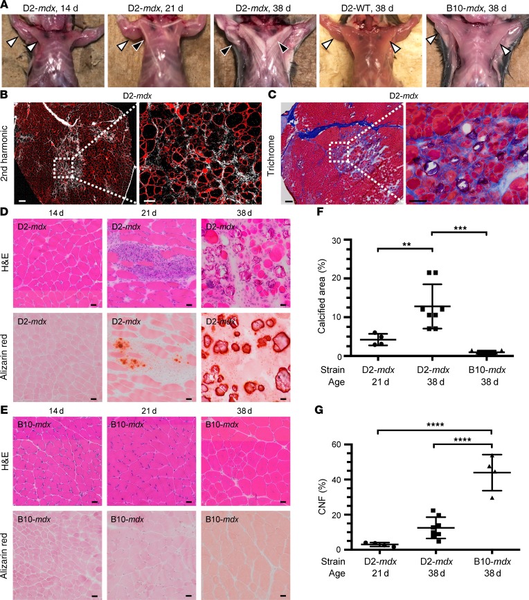
Figure 2. Muscles from D2-mdx mice show evidence of muscle degeneration and calcification starting at the onset of disease.
(A) Macroscopic images of 14-, 21-, and 38-day-old D2-mdx mice, 38-day-old D2-WT mice, and 38-day-old B10-mdx mice. D2-mdx mice show development of conspicuous pale stripes in the pectoral muscles between 21 and 38 days of age accompanied by similar changes in other muscles (e.g., triceps, black arrowhead) corresponding to areas of calcification identified in sections by alizarin red staining (D). No abnormal changes were seen in either D2-WT or B10-mdx mice (white arrowhead). (B and C) Signs of endomysial fibrosis in and around areas of degeneration in the triceps muscle were identified by second harmonic generation (B) and trichrome stain (C) in muscles of D2-mdx mice at 38 days of age. (D and E) H&E and alizarin red staining of triceps muscle sections from D2-mdx and B10-mdx mice at 14, 21, and 38 days of age. (F and G) Quantification of the percentage of calcified area and centrally nucleated fibers (CNFs) in triceps muscles from B10-mdx and D2-mdx at 21 days and 38 days old. (F) Quantification of percentage of calcification from 21-day-old D2-mdx mice (n = 4), 38-day-old D2-mdx mice (n = 7), and 38-day-old B10-mdx mice (n = 4). (G) Quantification of CNF percentage from 21-day-old D2-mdx mice (n = 4), 38-day-old D2-mdx mice (n = 8), and 38-day-old B10-mdx mice (n = 4). For F and G, data are expressed as mean ± SD. One-way ANOVA with Tukey’s post hoc comparison. **P < 0.01; ***P < 0.001; ****P < 0.0001. Scale bars: 50 μm (B and C), 20 μm (D and E).

