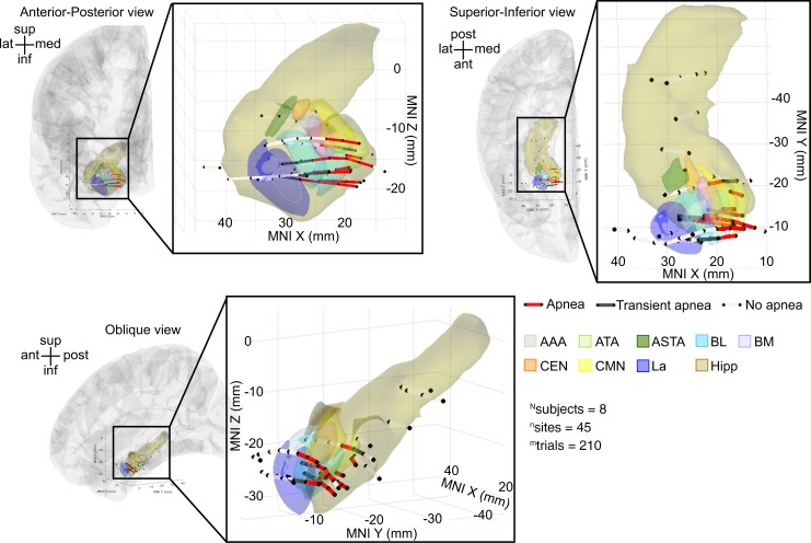
Figure 6. Across-subject analysis localized amygdala stimulation-induced apnea to the medial aspect of BL/BM and the ATA/CMN.
Anterior-posterior, superior-inferior, and oblique views of all stimulated electrode pairs in the amygdala and hippocampus across subjects plotted in a common coordinate system (MNI). Electrode contact pairs that produced apnea were located in the medial BL/BM and ATA/CMN. Electrode contact pairs that produced transient apnea were typically located just lateral or adjacent to this medial BL/BM region. Electrode contact pairs that failed to induce apnea were located in La, outside the amygdala, or in the hippocampus. Electrode pairs that induced apnea are denoted by dark red lines; those that produced transient apnea are depicted in dark gray lines; sites that did not induce apnea are depicted in light gray. For clarity, electrode sites on the lateral convexity, in the cingulate gyrus, and gyrus rectus are not shown; no sites omitted from this figure demonstrated effects of electrical stimulation on breathing. See Supplemental Table 1 for a list of MNI coordinates and the respiratory effect for each contact pair. Nuclei are color-coded with the same convention as in Figure 1B. Electrode contacts may appear outside of the template brain due to anatomical variation across subjects relative to the MNI coordinate system. All electrode contacts were plotted in the right hemisphere for simplicity, because no differences were observed between right and left amygdala stimulation.

