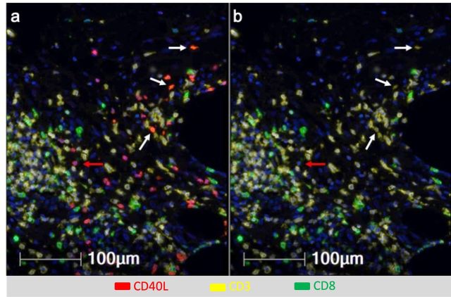
Figure 2.
Multiparameter immunofluorescence histology of the vaccine site microenvironment postvaccination with peptides in IFA and pICLC showing co-expression of CD40L and CD3 but not CD8. Selected markers include Dapi (blue), CD40L (red), CD3 (yellow), CD8 (green). (A) Includes CD40L and (B) excludes CD40L. White arrows depict CD3+CD8neg T cells co-expressing CD40L and CD3 but not CD8. Red arrows depict cells expressing CD40L but not CD3 or CD8.

