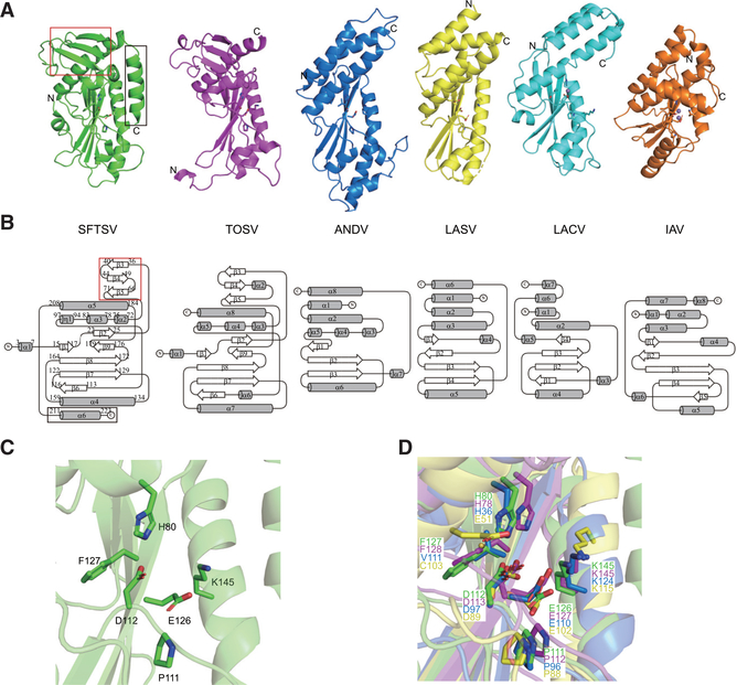
Figure 2. The Structural Fold of sNSV Endonuclease Domains Is Conserved among sNSVs.
(A) Comparisons of the endonuclease structures of SFTSV (green, PDB: 6NTV), TOSV (purple, PDB: 6QW0), ANDV (blue, PDB: 5HSB), LASV (yellow, PDB: 5J1P), LACV (cyan, PDB: 2XI5), and IAV (orange, PDB: 5DES). Manganese ions are shown as purple spheres in TOSV, ANDV, LASV, LACV, and IAV structures. Key catalytic residues are shown as sticks.
(B) Topological diagrams generated by TopDraw of endonuclease domains shown in (A) with the common core α helices represented as gray cylinders and the β strands as white arrows. The SFTSV L endonuclease domain contains a unique β sheet insertion at the N terminus (red box) and an extra C-terminal helix (black box).
(C) Close-up view of the active site of the SFTSV L endonuclease domain. Key catalytic residues are shown as sticks.
(D) Close-up view of superimposed endonuclease structures of SFTSV (green), TOSV (purple), ANDV (blue), and LASV (yellow). Key catalytic residues are shown as sticks.

