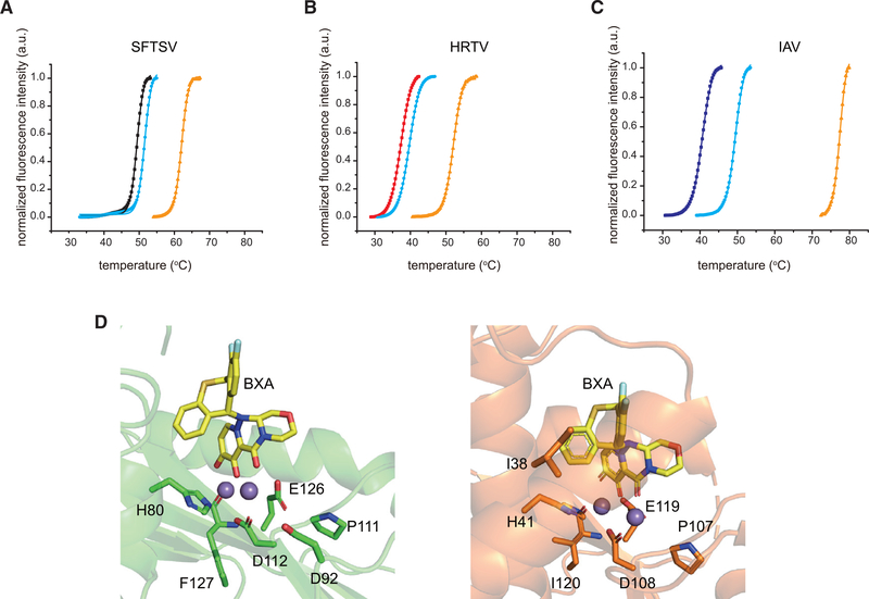
Figure 6. Inhibitor binding to SFTSV L endonuclease stabilizers its endonuclease domain.
Thermal stability was measured in the presence of 10 mM EGTA for: (A) SFTSV L 1–231 aa (black), (B) HRTV L 1–231 aa (red), or (C) IAV PA 1–192 aa (blue). Thermal stability for each viral endonuclease was also measured in the presence of 2 mM MnCl2 (cyan) and 2 mM MnCl2 with 0.2 mM BXA (orange). The lines represent the non-linear fits of the normalized fluorescence curves to Boltzmann equation. (D) Left, docking simulation of two manganese ions (purple) and BXA (yellow) with SFTSV L 1–231 aa (green). Key residues are shown in stick representation. Right, Structure of BXA bound to IAV PA (PDB 6FS6) is shown for comparison.

