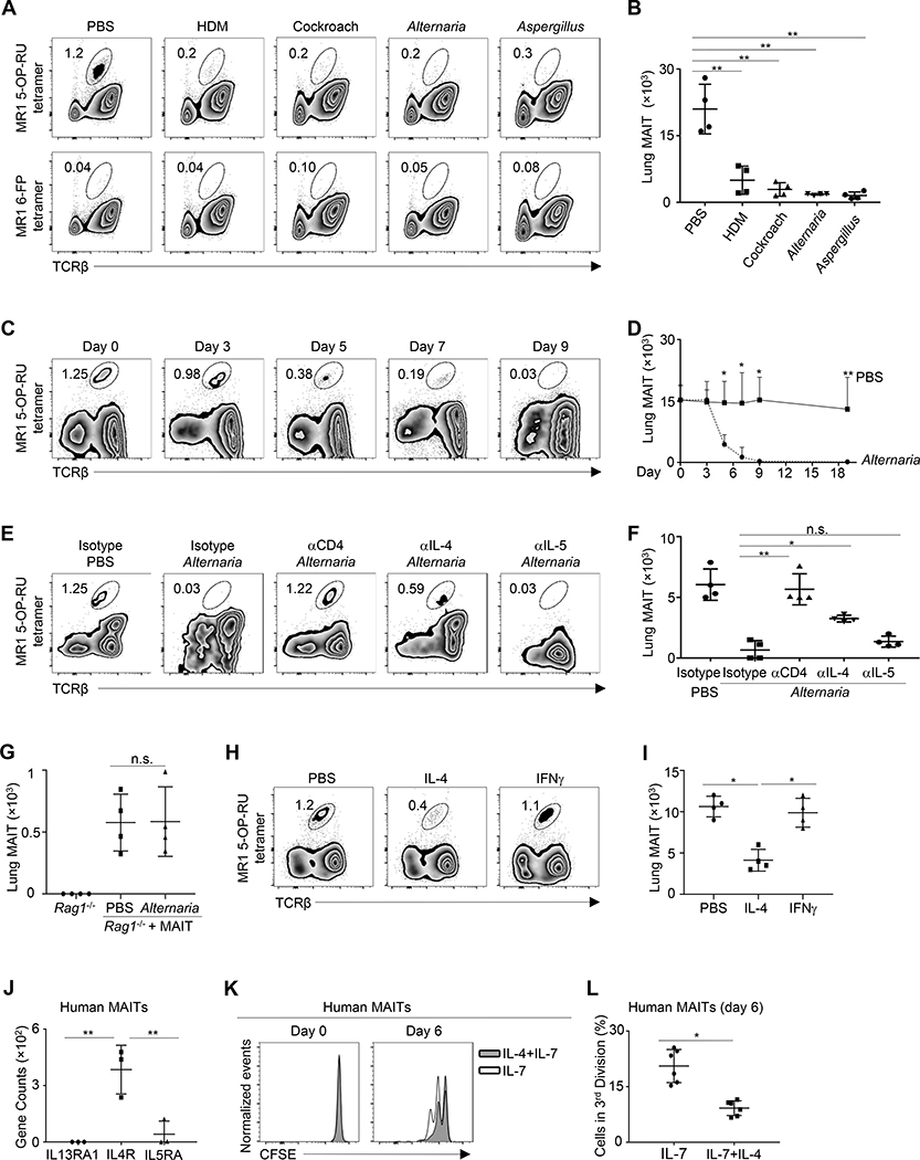
Figure 1. MAIT cells are diminished during allergic airway inflammation.
A, Lung MAIT cells in C57BL/6 mice challenged with the indicated allergen extracts every other day for 19 days. B, Numbers of lung MAIT cells in allergen-challenged mice. C, Lung MAIT cells in mice challenged with Alternaria extracts every other day for the indicated time points. D, Numbers of lung MAIT cells in Alternaria-challenged mice. E, Lung MAIT cells in mice challenged with Alternaria extracts and treated with the indicated antibodies every other day for 7 days. F, Numbers of lung MAIT cells in mice challenged with Alternaria and treated with the indicated antibodies. G, Numbers of lung MAIT cells in Rag1−/− mice that received MAIT cell transfer and challenged with Alternaria or PBS every other day for 7 days. H, Lung MAIT cells in mice treated with IL-4 or IFNγ daily for 7 days. I. Numbers of MAIT cells in cytokine-trmice treated with the indicated cytokines. J. Gene expression by RNA-seq. K, CFSE levels in MAIT cells before and after culturing with IL-7, or together with IL-4. L, Percentage of MAIT cells in the third division at day 6 of culture. N= 4 mice or 6 human samples per group, 2–3 independent experiments. *p < 0.05; **p < 0.01.

