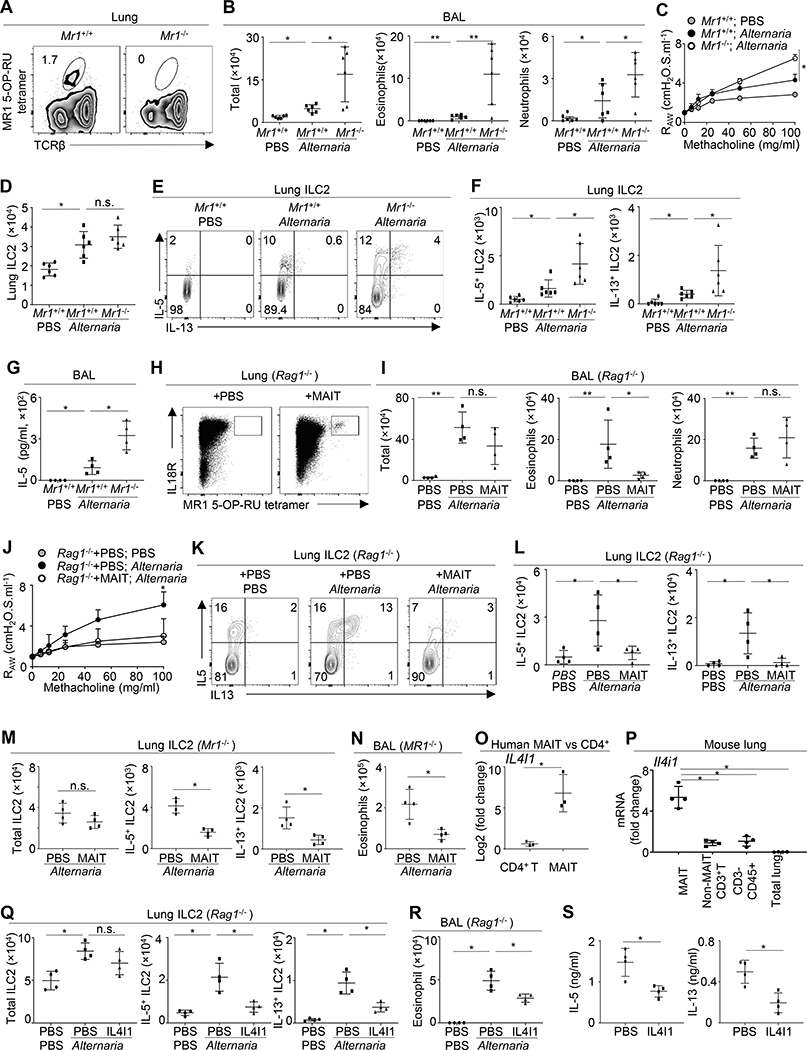
Figure 2. MAIT cells restrict Alternaria-induced airway inflammation and hyperresponsiveness.
A, Lung MAIT cells in Mr1+/+ and Mr1−/− mice. B, Bronchoalveolar lavage (BAL) cells in mice challenged with PBS or Alternaria for 3 days. C, FlexiVent analysis. D, Numbers of lung ILC2. E. Cytokine expression by ILC2. F, Numbers of IL-5+ or IL-13+ ILC2. G, BAL IL-5 concentrations. H, Lung MAIT cells in Rag1−/− mice with or without MAIT cell transfer. I, BAL cell numbers in Rag1−/− mice challenged with Alternaria or PBS, with or without MAIT cell transfer. J, FlexiVent analysis. K, Cytokine expression by lung ILC2. L, Numbers of IL-5+ or IL-13+ ILC2. M, Numbers of ILC2 in Mr1−/− mice challenged with Alternaria, with or without MAIT cell transfer. N, BAL eosinophilic numbers. O, IL4I1 expression in human blood CD4+ T cell and MAIT cells. P, Il4i1 expression in lung MAIT cells, non-MAIT T cells, non-T immune cells, and the whole lung tissue of naïve mice. Q, Numbers of ILC2 in Rag1−/− mice challenged with PBS or Alternaria, with or without IL4I1 treatment. R, BAL eosinophilic numbers. S, IL-5 and IL-13 production in mouse lung ILC2 cultured with or without IL4I1. N= 3–6 mice per group, 3–4 independent experiments. *p < 0.05; **p < 0.01.

