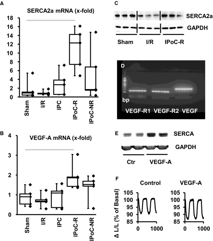
Figure 3.

Effect of VEGF‐A on SERCA expression and functional improvement. A + B, mRNA expression of SERCA2a (A) and VEGF‐A (B) in the five groups expressed as box and whisker plots and individual data points. C, Immunoblot of SERCA2a expression and a loading control (GAPDH). D, mRNA expression of VEGF receptor 1 and 2 and VEGF‐A in cardiomyocyte preparations. E, Immunoblot indicating protein levels of SERCA2a 24 h after incubation with VEGF‐A (50 µg/mL); F, Effect of VEGF‐A on load‐free cell shortening expressed as shortening amplitude · 100/ diastolic cell length (ΔL/L)
