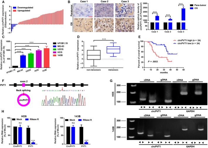
FIGURE 1.

CircPVT1 is up‐regulated in OS and correlated with poor outcomes. A, CircPVT1 expression in 48 paired OS tissue samples was qualified by using a qRT‐PCR analysis via applying of log2 (2−△△Ct) method. B, CircPVT1 expression in OS tissue samples and paired para‐tumour tissue samples was determined by an ISH assay. Scale bar, 50 µm; magnification, 40×. C, qRT‐PCR was used to measure the expression of circPVT1 in 4 OS cell lines MG‐63, U2OS, HOS and 143B and in a normal human osteoblast cell line hFOB1.19. D, Expression of circPVT1 was higher in patients with lymph node metastasis (N1 and N2, than that in patients without lymph node metastasis (N0). E, Association of circPVT1 expression with overall survival analysis of 48 OS patients, detected by a Kaplan‐Meier analysis, P = .0053. F, Sanger sequencing was used to verify the head‐to‐tail splicing of circPVT1. G, RT‐PCR validated the existence of circPVT1 in HOS and 143B cell lines. CircPVT1 in cDNA, instead of genomic DNA, was amplified by divergent primers. GAPDH was used as a negative control. H, Expression of circPVT1 and PVT1 in HOS and 143B cells were determined by RT‐PCR or qRT‐PCR with or without the effect of RNase R. Data were shown as mean ± SD from three independent experiments. ***P < .001 compared to controls
