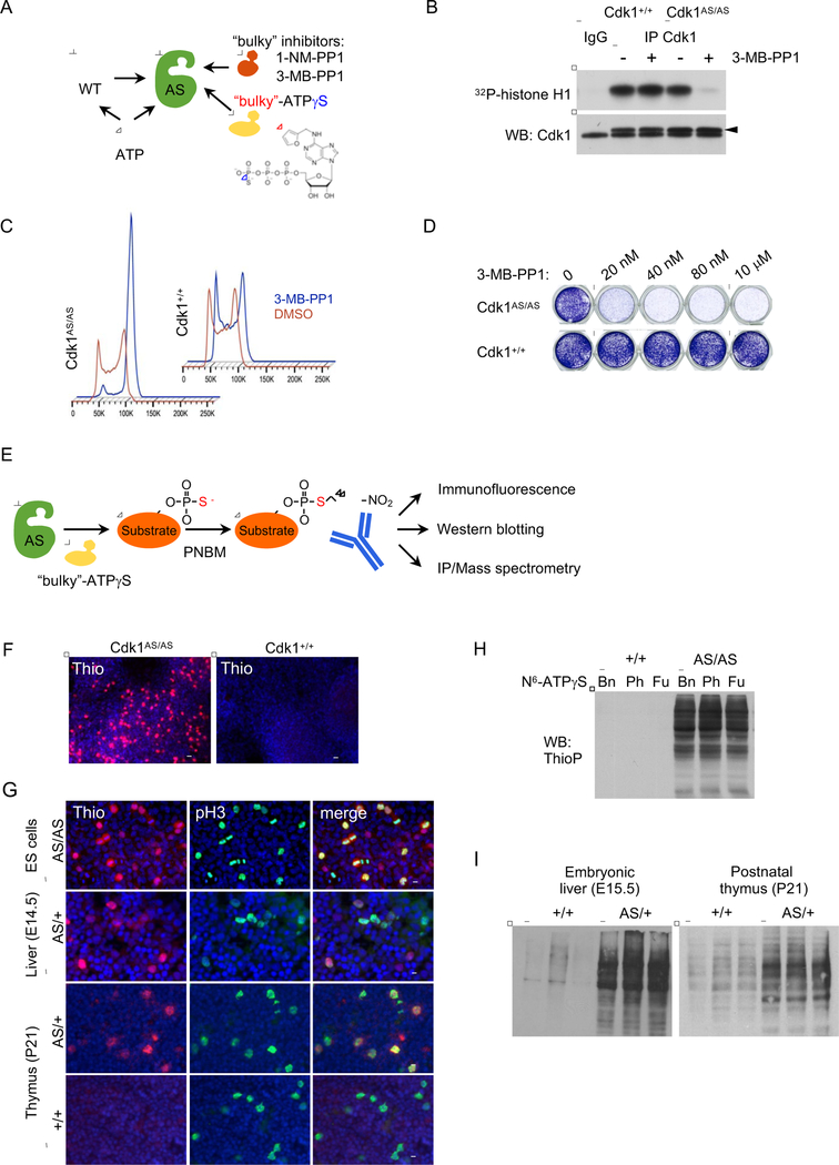
Figure 1. Analyses of Embryonic Stem Cells and Organs Expressing Analog-Sensitive Cdk1.
(A) Diagram illustrating different experimental approaches using analog-sensitive kinases.
(B) Endogenous Cdk1 was immunoprecipitated from Cdk1+/+ or Cdk1AS/AS ES cells and subjected to in vitro kinase reactions with histone H1 as a substrate. For control, immunoprecipitates obtained with normal IgG (IgG) were used for kinase reactions. Lower panel, immunoblots were probed with antibodies against Cdk1 (arrowhead points to the band corresponding to Cdk1). +3-MB-PP1 denotes addition of 3-MB-PP1.
(C) Cdk1+/+ or Cdk1AS/AS ES cells were cultured for 18 h in the presence of vehicle (DMSO), or 3-MB-PP1, stained with propidium iodide and analyzed by flow cytometry.
(D) Cdk1+/+ or Cdk1AS/AS ES cells were cultured for 4 d in the presence of the indicated concentrations of 3-MB-PP1, and stained with crystal violet. Vertical lines mark places where the image was combined from non-adjacent wells.
(E) Experimental outline of using Cdk1AS/AS cells for identification of substrates. PNBM, p-nitrobenzyl mesylate used to alkylate proteins in order to generate epitopes for an anti-thiophosphate ester antibody.
(F) In vitro cultured Cdk1AS/AS or Cdk1+/+ ES cells were permeabilized by treatment with digitonin, and supplemented with N6-substituted bulky ATP analog, N6-furfuryl-ATPγS for 20 min. Cells were then alkylated with PNBM, and thio-labeled substrates visualized by immunostaining with an anti-thiophosphate ester antibody. Scale bar, 50 μm.
(G) Detection of Cdk1 substrates in in vitro cultured Cdk1AS/AS ES cells or in frozen sections prepared from embryonic day 14.5 fetal livers or postnatal day 21 thymuses isolated from Cdk1AS/+ (AS/+) or control Cdk1+/+ mice (+/+). Staining for phospho-histone H3 (pH3) was used to detect mitotic cells. Scale bar, 20 μm
(H)In vitro cultured ES cells were permeabilized and supplemented with one of N6-substituted bulky ATPγS analogs, N6-benzyl-ATPγS (Bn), N6-phenylethyl-ATPγS (Ph), or N6-furfuryl-ATPγS (Fu) for 20 min. Proteins were then analyzed by western blotting with an anti-thiophosphate ester antibody to detect thio-phosphorylated proteins (representing Cdk1 phosphorylation substrates).
(I) Protein lysates were prepared from the indicated organs of Cdk1AS/+ mice and supplemented for 20 min with N6-furfuryl-ATPγS (to allow labeling of direct Cdk1 substrates). Lysates were then immunoblotted using an anti-thiophosphate ester antibody to detect thio-phosphorylated proteins.
See also Figure S1.

