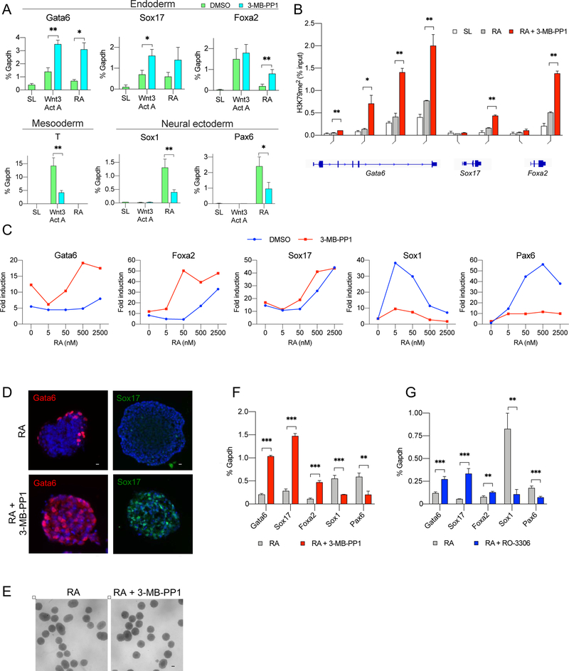
Figure 7. Inhibition of Cdk1 Promotes Endodermal Differentiation.
(A) RT-qPCR analysis of the levels of the indicated transcripts in Cdk1AS/AS ES cells cultured in stem cell medium (SL) or switched to differentiation medium containing Wnt3a and activin A or retinoic acid (RA), with vehicle (DMSO) or 3-MB-PP1. T represents a mesodermal marker, Sox1 and Pax6 markers of neural ectoderm. Mean values ±SD from 3 independent experiments. *, p<0.05; **, p<0.01 (unpaired t-test).
(B) Targeted ChIP – qPCR analysis to assess enrichment of H3K79me2 marks on the promoter and gene body regions of the Gata6, Sox17 and Foxa2 genes. Cdk1AS/AS ES cells were cultured in stem cell medium (SL), or switched to differentiation medium containing retinoic acid (RA), or retinoic acid plus 3-MB-PP1, and subjected to ChIP with an anti-H3K79me2 antibody, followed by PCR amplification of the indicated gene segments. Mean values ±SD from 2 independent experiments. *, p<0.05; **, p<0.01 (multiple t-test with Benjamini’s correction).
(C) Cdk1AS/AS ES cells were cultured for 2 days in a serum- and LIF-free medium to dissolve pluripotency, followed by 24 h culture in the presence of the indicated concentrations of retinoic acid with DMSO or 3-MB-PP1. Expression of transcripts encoding endodermal (Gata6, Foxa2 and Sox17) and ectodermal (Sox1, Pax6) markers was gauged by RT-qPCR. Shown is fold induction relative to expression levels in cells growing in stem cell SL medium.
(D) Cdk1AS/ASES cells were allowed to form embryoid bodies for 2 days and then cultured in the presence of retinoic acid (RA, upper row) or with retinoic acid plus 3-MB-PP1 (lower row) for 24 h. Embryoid bodies were then cryosectioned and stained with antibodies against Gata6 and Sox17. DAPI was used to visualize cell nuclei. Scale bar, 20 μm.
(E) Bright field images of embryoid bodies cultured as above. Scale bar, 100 μm. Note that treatment with 3-MB-PP1 did not have any gross effects on morphology of embryoid bodies.
(F) Transcript levels of the indicated markers in embryoid bodies grown and treated as in D.
(G) Embryoid bodies derived from wild-type ES cells were generated as in D and F and supplemented with retinoic acid alone, or retinoic acid plus a chemical inhibitor of Cdk1, RO-3306. The expression of the indicated markers was analyzed by RT-qPCR. In (F) and (G), mean values ±SD from 3 biological replicates. **, p<0.01; **, p<0.001 (multiple t-test with Benjamini’s correction).

