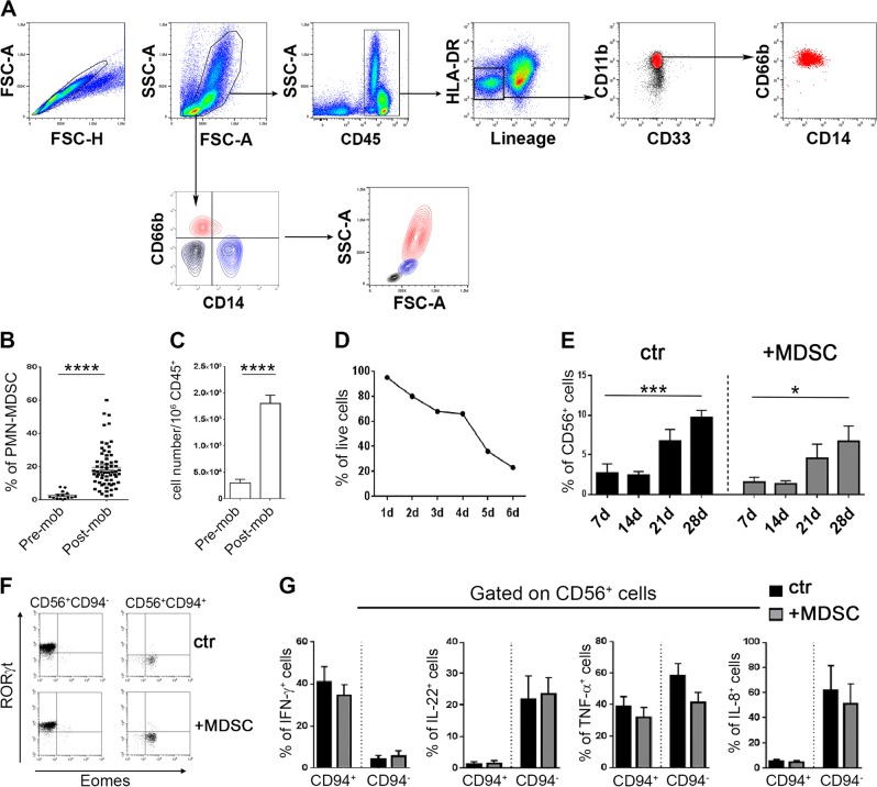
Fig. 1.
Presence of PMN-MDSC in the apheresis of G-CSF-mobilized donors and their effect on NK/ILC differentiation from HSC. a–c Mononuclear cells present in the apheresis were analyzed by flow-cytometry for the expression of specific markers allowing the identification of MDSC subsets. a Gating strategy adopted in one representative experiment out of 70 performed. b Percentages and c absolute numbers of PMN-MDSC (CD66b+ cells) in different donors before (pre) and after (post) mobilization (mob). d Percentages of PMN-MDSC viable cells at different time points. e–g In these experiments, CD34+ HSC were isolated from the apheresis of G-CSF-mobilized donors and cultured with cytokine-medium either in the absence (ctr) or in the presence (+MDSC) of PMN-MDSC (at 1/1 ratio) and analyzed by flow-cytometry for: e percentages ± SEM of CD56+ cells at different time points (n = 7); f RORγt and Eomes transcription factor expression in gated CD56+CD94+ cells (NK cells) and CD56+CD94− (ILC3) at day 40 of culture. One representative experiment out of seven performed; g expression of informative intracellular cytokines (IFN-γ, IL-22, TNF-α, and IL-8) at day 40 in CD56+CD94+ or CD56+CD94− cells after stimulation with PMA+Ionomycine+IL-23. Percentage ± SEM (n = 3–7). A p value ≤ 0.05 was considered statistically significant. *p ≤ 0 .05; **p ≤ 0 .01; *** p ≤ 0.001; **** p ≤ 0.0001; ns = not significant. Where not indicated, the data were not statistically significant

