Fig. 2.
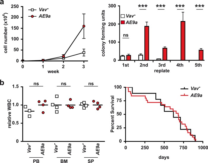
AE9a-expressing bone marrow cells exhibit enhanced stem cell characteristics but do not initiate leukemogenesis. a, left, AE9a-expressing cells show enhanced proliferation capacity. Proliferation potential of Lineage−, cKit+, GFP+ cells from 12 weeks old AE9a mice (red dots) and Lineage−, cKit+ cells isolated from Cre-negative littermate controls (Vav−, white squares) in suspension culture was estimated by cell number counts taken in seven days intervals over 3 weeks. MW ± SD, n = 2. a, right, AE9a-expressing cells show significant self-renewal capacity. Colony-forming potential of Lineage−, cKit+, GFP+ cells from 12 weeks old AE9a mice (red bars) and Lineage−, cKit+ cells isolated from Vav1− littermate controls (white bars) was measured by serial replating on semi-solid methylcellulose medium in seven days intervals over 5 weeks. MW ± SD (error bars) of the colony-forming units of triplicates from one representative experiment with n = 2 mice/group is shown. b, left, White blood cell counts (WBC) are not altered in 16 weeks old AE9a mice (red dots) compared to Vav− littermate controls (white squares). Individual and mean values of peripheral blood (PB), bone marrow (BM) and spleen (SP) from n = 4 mice/group are shown relative to the mean of the respective Vav− group. (b, right) AE9a expression in the hematopoietic compartment does not influence survival of mice. Kaplan–Meier plot illustrating that survival in AE9a mice (red line, mean survival 580 days, n = 25) is not altered compared to Vav− control mice (black line, mean survival 559 days, n = 9). ns, not significant; ***p < 0.001

