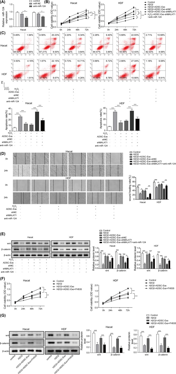
Figure 6. ADSC-Exos containing MALAT1 mediates H2O2 induced wound healing by targeting miR-124 through activating Wnt/β-catenin pathway.
(A) The expression level of miR-124 was detected by qRT-PCR after transfected with anti-miR-124. (B) CCK-8 assay was performed to assess the cell proliferation of HaCaT and HDF cells treated with H2O2, H2O2+ADSC-Exo, H2O2+ADSC-Exo-shNC, H2O2+ADSC-Exo-shMALAT1, H2O2+ADSC-Exo-shMALAT1+anti-miR-124. (C) Flow cytometry was subjected to evaluate cell apoptosis of HaCaT and HDF cells treated with H2O2, H2O2+ADSC-Exo, H2O2+ADSC-Exo-shNC, H2O2+ADSC-Exo-shMALAT1, H2O2+ADSC-Exo-shMALAT1+anti-miR-124. (D) Migration of HaCaT and HDF cells treated with H2O2, H2O2+ADSC-Exo, H2O2+ADSC-Exo-shNC, H2O2+ADSC-Exo-shMALAT1, H2O2+ADSC-Exo-shMALAT1+anti-miR-124 were analyzed by the scratch wound healing assay. (E) The expression of Wnt/β-catenin signals were determined by western blot. (F) Cell proliferation of HaCaT and HDF cells treated with FH535 were evaluated by CCK-8 assay. (G) Wnt/β-catenin signal pathway of HaCaT and HDF cells treated with H2O2, H2O2+ADSC-Exo or H2O2+ADSC-Exo+FH535 were monitored by western blot. *P<0.05, **P<0.01, ***P<0.001.

