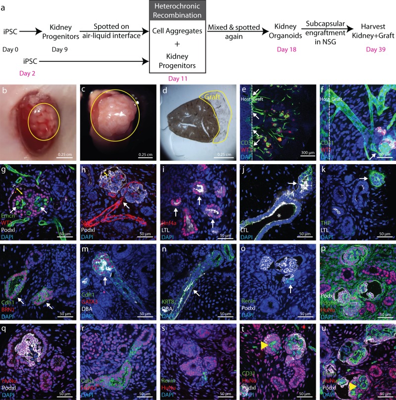
Fig. 4. In vivo maturation of kidney organoids.
a Schematic diagrams showing generation of kidney organoids and their subcapsular engraftment in mice kidney. b Image showing kidney organoids immediately after engraftment. c Three weeks after engraftment organoids grows extensively in a big tissue. d Stereo microscope image showing vibratome section of mouse kidney with graft for further analysis. e Low magnification immunofluorescence image showing CD31+ endothelial network coming from host kidney widespread in the graft. f High magnification immunofluorescence image showing these CD31+ endothelial networks are connecting with Podxl+ WT1+ presumptive glomeruli. g and h Immunofluorescence images showing characteristic morphology of glomerular structure including Podxl+ WT1+ podocytes, Emcn+ capillary tuft and bowman’s space (yellow double arrow head). These glomerular structures also shows direct connection with SMA+ vascular network. i Immunofluorescence image showing Hnf4a+ and LTL+ proximal tubule and j this LTL+ Cdh1− proximal tubule segmented in LTL− Cdh1+ distal tubule with lumen (white arrow head). k Immunofluorescence image showing THP+ loop of Henle and l Brn1+ Cdh1+ distal tubule. m and n Immunofluorescence image showing Cdh1+ GATA3+ DBA+ connecting tubule or collecting duct connected with Cdh1+ GATA3− DBA− distal segment of nephron. In addition, immunofluorescence images showing KRT+ DBA+ extreme distal segment or collecting duct. o Further, immunofluorescence image showing Renin+ juxtaglomerular cells at the base of Podxl+ glomerular structure. We did not observe Renin+ cells in organoid before engraftment which is another confirmation of organoid maturation in vivo. p–s Immunofluorescence image showing Pdgfrβ+ mesangial cells surrounded by Podxl+ Podocytes inside presumptive glomerular structures. Further immunofluorescence images showing differentiated Pdgfrβ+ mesangial or interstitial cells, Podxl+ podocytes, Cdh1+ epithelial structures and Renin+ cells were derived from human cells (HuNu+) but t–u Emcn+/CD31+ endothelial cells were derived from mouse (HuNu−, yellow arrow head). Images shown here are representative and were derived from n = 6 NSG mice.

