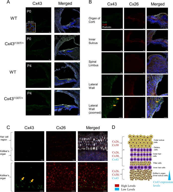
Fig. 2. Low but detectable amounts of Cx43 are expressed in organotypic cochlear cultures.
a 3D reconstruction of consecutively stacked confocal images of cochleae obtained from Cx43I130T/+ mutant mice and their WT littermates (P0 and P4) apical regions. Insert = HEI-OC1 cells. b Higher-magnification images of specific cochlear regions of WT P4 cochleae. c Stacked confocal images of organotypic cochlear cultures from WT mice of apical regions. d Schematic diagram representing the localization and relative expression levels of cochlear connexins in organotypic cochlear cultures. Cx43 is denoted in green, Cx26 is in red, phalloidin is in white, and Hoechst nuclear stain is in blue. Orange arrowheads indicate areas of gap junction plaques. Scale bars = 10 μm.

